Foetal testis, light micrograph
Bildnummer 13686898
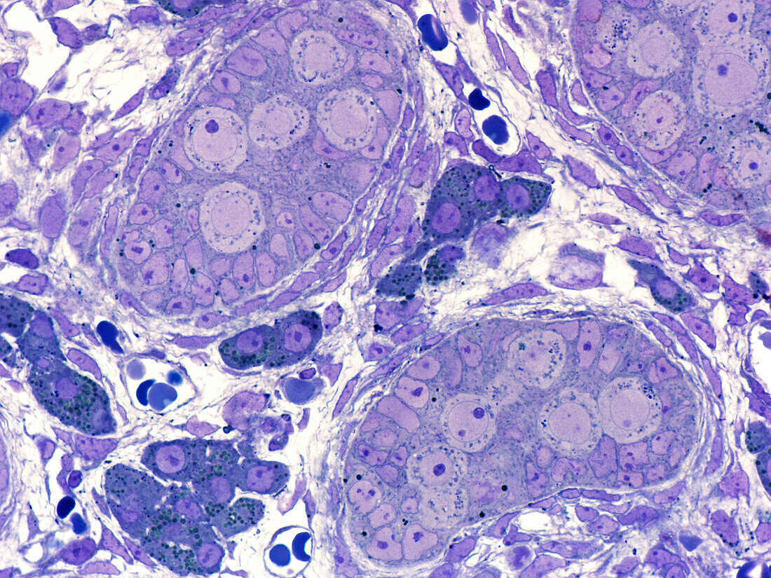
| Foetal testis, light micrograph. Seminiferous cords contain pale-staining round spermatogonia and numerous densely packed immature Sertoli cells of irregular shape. Cords are bordered by several layers of elongated mesenchymal cells and fibroblasts. In the interstitial tissue between the cords are clusters of Leydig cells (deep blue) secreting high levels of androgens that stimulate testis development. Epoxy resin section, Toluidine blue stain. Magnification: x900 when width printed at 10cm. | |
| Lizenzart: | Lizenzpflichtig |
| Credit: | Science Photo Library / Microscape |
| Bildgröße: | 4843 px × 3632 px |
| Modell-Rechte: | nicht erforderlich |
| Eigentums-Rechte: | nicht erforderlich |
| Restrictions: | - |
Preise für dieses Bild ab 15 €
Universitäten & Organisationen
(Informationsmaterial Digital, Informationsmaterial Print, Lehrmaterial Digital etc.)
ab 15 €
Redaktionell
(Bücher, Bücher: Sach- und Fachliteratur, Digitale Medien (redaktionell) etc.)
ab 30 €
Werbung
(Anzeigen, Aussenwerbung, Digitale Medien, Fernsehwerbung, Karten, Werbemittel, Zeitschriften etc.)
ab 55 €
Handelsprodukte
(bedruckte Textilie, Kalender, Postkarte, Grußkarte, Verpackung etc.)
ab 75 €
Pauschalpreise
Rechtepakete für die unbeschränkte Bildnutzung in Print oder Online
ab 495 €