Foetal testis, light micrograph
Bildnummer 13686893
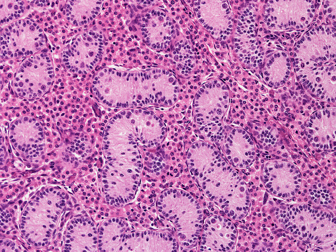
| Foetal testis, light micrograph. The foetal testis shows seminiferous cords cut in various planes surrounded by an abundant population of Leydig cells that secrete androgens. The cords contain Sertoli cells arranged around the periphery with lesser numbers of round germ cells/spermatogonia in the central regions. In postnatal life spermatogonia activate to start spermatogenesis with the cords greatly enlarging in diameter and length to form seminiferous tubules. The foetal Leydig cell numbers regress after birth and a new population of adult-type Leydig cells develop. Paraffin section, haematoxylin and eosin stain. Magnification: x230 when width printed at 10cm. | |
| Lizenzart: | Lizenzpflichtig |
| Credit: | Science Photo Library / Microscape |
| Bildgröße: | 4724 px × 3543 px |
| Modell-Rechte: | nicht erforderlich |
| Eigentums-Rechte: | nicht erforderlich |
| Restrictions: | - |
Preise für dieses Bild ab 15 €
Universitäten & Organisationen
(Informationsmaterial Digital, Informationsmaterial Print, Lehrmaterial Digital etc.)
ab 15 €
Redaktionell
(Bücher, Bücher: Sach- und Fachliteratur, Digitale Medien (redaktionell) etc.)
ab 30 €
Werbung
(Anzeigen, Aussenwerbung, Digitale Medien, Fernsehwerbung, Karten, Werbemittel, Zeitschriften etc.)
ab 55 €
Handelsprodukte
(bedruckte Textilie, Kalender, Postkarte, Grußkarte, Verpackung etc.)
ab 75 €
Pauschalpreise
Rechtepakete für die unbeschränkte Bildnutzung in Print oder Online
ab 495 €