Cerebellum tissue, light micrograph
Bildnummer 13673834
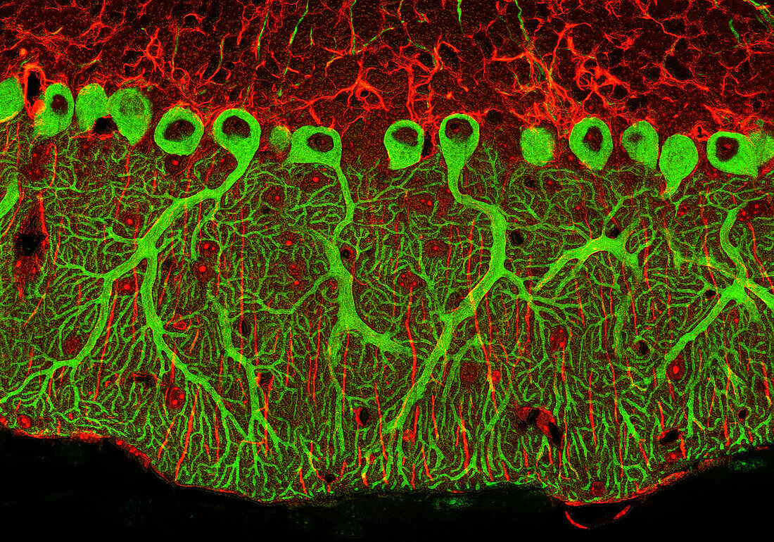
| Cerebellum tissue. 2-photon fluorescent micrograph of a section through the cerebellum of the brain. Purkinje cells, a type of neuron (nerve cell), are bight green due to staining with anti-IP3 receptor antibodies and quantum dot 565. They consist of a flask-shaped cell body with many branching processes (dendrites) that receive impulses from other cells. Purkinje cells form the junction between the granular (red) and molecular (green) layers of the grey matter (cortex) of the cerebellum. Glial cells in the granular layer are stained red with anti-GFAP (glial fibrillary acidic protein) with Quantum dot 655 to The cerebellum controls balance, posture and muscle coordination. Quantum dots are nanocrystals of semiconductor material that emit photons when excited. The size of the quantum dot can be tuned during manufacturing to produce a certain wavelength of photon emission. | |
| Lizenzart: | Lizenzpflichtig |
| Credit: | Science Photo Library / Deerinck, Thomas / NCMIR |
| Bildgröße: | 3900 px × 2730 px |
| Modell-Rechte: | nicht erforderlich |
| Eigentums-Rechte: | nicht erforderlich |
| Restrictions: | - |
Preise für dieses Bild ab 15 €
Universitäten & Organisationen
(Informationsmaterial Digital, Informationsmaterial Print, Lehrmaterial Digital etc.)
ab 15 €
Redaktionell
(Bücher, Bücher: Sach- und Fachliteratur, Digitale Medien (redaktionell) etc.)
ab 30 €
Werbung
(Anzeigen, Aussenwerbung, Digitale Medien, Fernsehwerbung, Karten, Werbemittel, Zeitschriften etc.)
ab 55 €
Handelsprodukte
(bedruckte Textilie, Kalender, Postkarte, Grußkarte, Verpackung etc.)
ab 75 €
Pauschalpreise
Rechtepakete für die unbeschränkte Bildnutzung in Print oder Online
ab 495 €
Keywords
- Anatomie,
- anatomisch,
- Antikörper,
- Biologie,
- biologisch,
- Dendrit,
- Fluoreszenz,
- fluoreszierend,
- Gehirn,
- gesund,
- Halbleiter,
- Kleinhirn,
- Lichtmikroskop,
- lichtmikroskopische Aufnahme,
- Mausgewebe,
- Mikrofotografie,
- molekulare Schicht,
- Nano,
- Nanotechnologie,
- Nervenzelle,
- Neuron,
- Neuronen,
- Neurowissenschaften,
- normal,
- Optik,
- Sektion,
- sektioniert,
- Zellen,
- zentrales Nervensystem