Human thymus, light micrograph
Bildnummer 13673502
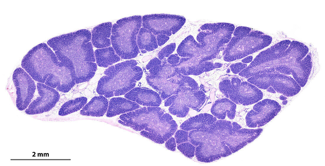
| Light micrograph of a human thymus gland stained with haematoxylin and eosin. The thymic parenchyma is composed of small lobules separated by adipose (fat) tissue showing a dark peripheral cortex and a central clear medulla. The peripheral cortex appears more stained, due to the high density of T-lymphocyte precursor cells. In the paler centre of each lobule there are many Hassall's corpuscles. The scale bar corresponds to 2 mm. | |
| Lizenzart: | Lizenzpflichtig |
| Credit: | Science Photo Library / JOSE CALVO |
| Bildgröße: | 4878 px × 2500 px |
| Modell-Rechte: | nicht erforderlich |
| Eigentums-Rechte: | nicht erforderlich |
| Restrictions: | - |
Preise für dieses Bild ab 15 €
Universitäten & Organisationen
(Informationsmaterial Digital, Informationsmaterial Print, Lehrmaterial Digital etc.)
ab 15 €
Redaktionell
(Bücher, Bücher: Sach- und Fachliteratur, Digitale Medien (redaktionell) etc.)
ab 30 €
Werbung
(Anzeigen, Aussenwerbung, Digitale Medien, Fernsehwerbung, Karten, Werbemittel, Zeitschriften etc.)
ab 55 €
Handelsprodukte
(bedruckte Textilie, Kalender, Postkarte, Grußkarte, Verpackung etc.)
ab 75 €
Pauschalpreise
Rechtepakete für die unbeschränkte Bildnutzung in Print oder Online
ab 495 €