Abdomen, illustration
Bildnummer 13672587
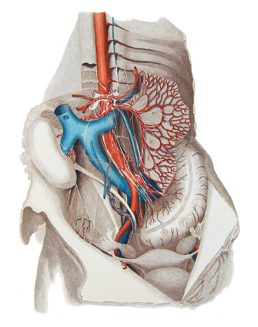
| Illustration of the neurovascular structures of the abdomen. The inferior vena cava (blue) and the right kidney are displaced to the right of the midline. Branches of the abdominal aorta (red) are shown, including the arterial arcades of the superior mesenteric artery passing through the cut mesentery, and the inferior mesenteric supplying the descending colon. The autonomic nerve plexuses of the celiac and superior mesenteric ganglia are clustered around the abdominal aorta. From Lizars, J. 1823 A system of anatomical plates of the human body. W.H. Lizars, Edinburgh. | |
| Lizenzart: | Lizenzpflichtig |
| Credit: | Science Photo Library / Microscape |
| Bildgröße: | 3971 px × 5028 px |
| Modell-Rechte: | nicht erforderlich |
| Eigentums-Rechte: | nicht erforderlich |
| Restrictions: | - |
Preise für dieses Bild ab 15 €
Universitäten & Organisationen
(Informationsmaterial Digital, Informationsmaterial Print, Lehrmaterial Digital etc.)
ab 15 €
Redaktionell
(Bücher, Bücher: Sach- und Fachliteratur, Digitale Medien (redaktionell) etc.)
ab 30 €
Werbung
(Anzeigen, Aussenwerbung, Digitale Medien, Fernsehwerbung, Karten, Werbemittel, Zeitschriften etc.)
ab 55 €
Handelsprodukte
(bedruckte Textilie, Kalender, Postkarte, Grußkarte, Verpackung etc.)
ab 75 €
Pauschalpreise
Rechtepakete für die unbeschränkte Bildnutzung in Print oder Online
ab 495 €