Thorax and abdomen, illustration
Bildnummer 13672577
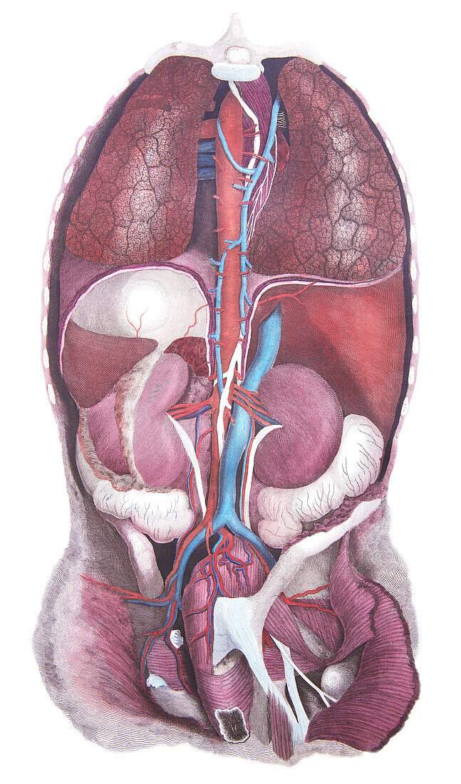
| Posterior view of the thoracic and abdominal viscera, illustration. Between the lungs is the thoracic aorta (red), azygos vein (blue) and thoracic duct (white). Below the cut diaphragm at left is the stomach (white) and spleen (deep red) and to the right is the vena cava (blue) and the liver (red). Renal arteries supply the kidneys from which the ureters (white) descend inferiorly. The abdominal aorta and inferior vena cava in the pelvic region are associated with their respective iliac vessels. The ascending colon (white) lies below the right kidney with the sigmoid colon (white) below the left kidney. From Lizars, J. 1823 A system of anatomical plates of the human body. W.H. Lizars, Edinburgh. | |
| Lizenzart: | Lizenzpflichtig |
| Credit: | Science Photo Library / Microscape |
| Bildgröße: | 3543 px × 6020 px |
| Modell-Rechte: | nicht erforderlich |
| Eigentums-Rechte: | nicht erforderlich |
| Restrictions: | - |
Preise für dieses Bild ab 15 €
Universitäten & Organisationen
(Informationsmaterial Digital, Informationsmaterial Print, Lehrmaterial Digital etc.)
ab 15 €
Redaktionell
(Bücher, Bücher: Sach- und Fachliteratur, Digitale Medien (redaktionell) etc.)
ab 30 €
Werbung
(Anzeigen, Aussenwerbung, Digitale Medien, Fernsehwerbung, Karten, Werbemittel, Zeitschriften etc.)
ab 55 €
Handelsprodukte
(bedruckte Textilie, Kalender, Postkarte, Grußkarte, Verpackung etc.)
ab 75 €
Pauschalpreise
Rechtepakete für die unbeschränkte Bildnutzung in Print oder Online
ab 495 €