Human cell in anaphase, light micrograph
Bildnummer 13672563
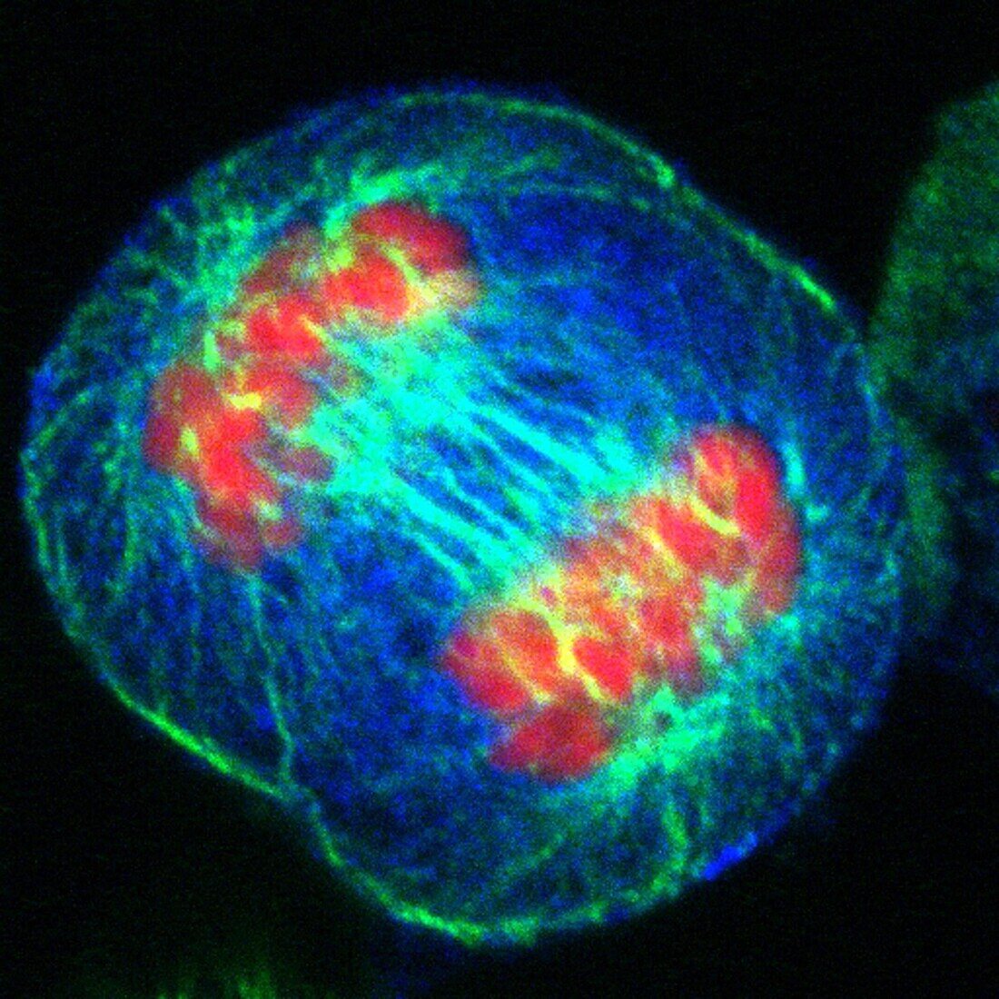
| Fluorescence light micrograph of a HeLa cell in anaphase showing the chromatin in red, the microtubules forming the spindle in green, and the cytoplasm in blue. The chromosomes are separating from each other and moving to the opposite poles of the cell. | |
| Lizenzart: | Lizenzpflichtig |
| Credit: | Science Photo Library / DR MATTHEW DANIELS |
| Bildgröße: | 3240 px × 3240 px |
| Modell-Rechte: | nicht erforderlich |
| Eigentums-Rechte: | nicht erforderlich |
| Restrictions: | - |
Preise für dieses Bild ab 15 €
Universitäten & Organisationen
(Informationsmaterial Digital, Informationsmaterial Print, Lehrmaterial Digital etc.)
ab 15 €
Redaktionell
(Bücher, Bücher: Sach- und Fachliteratur, Digitale Medien (redaktionell) etc.)
ab 30 €
Werbung
(Anzeigen, Aussenwerbung, Digitale Medien, Fernsehwerbung, Karten, Werbemittel, Zeitschriften etc.)
ab 55 €
Handelsprodukte
(bedruckte Textilie, Kalender, Postkarte, Grußkarte, Verpackung etc.)
ab 75 €
Pauschalpreise
Rechtepakete für die unbeschränkte Bildnutzung in Print oder Online
ab 495 €