Liver, light micrograph
Bildnummer 13633595
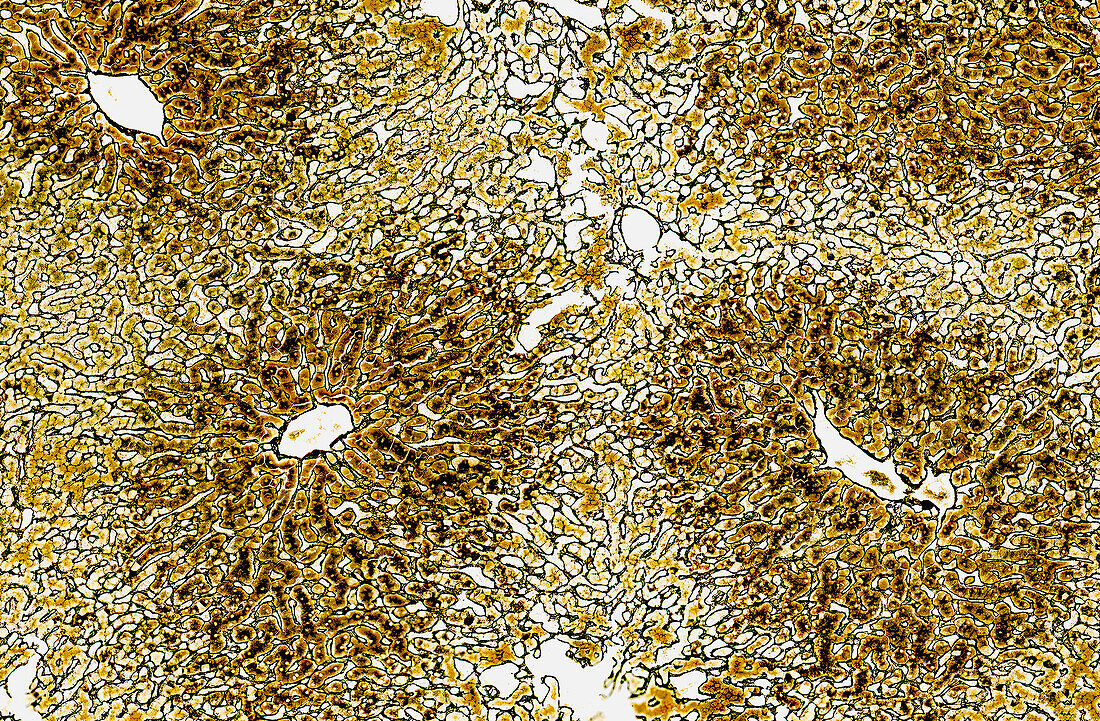
| Light micrograph of liver tissue showing radial cords of hepatocytes (orange-brown stain) arranged around a central vein. Between the rows of hepatocytes are many blood vessels termed hepatic sinusoids. Supporting the sinusoids is a connective tissue framework of reticular fibres consisting of type III collagen. The liver is thus heavily perfused by blood chiefly entering from the hepatic portal vein. Paraffin section, Gordon and Sweet's reticulin stain. Magnification: x55 when width printed at 10cm. | |
| Lizenzart: | Lizenzpflichtig |
| Credit: | Science Photo Library / Microscape |
| Bildgröße: | 5048 px × 3307 px |
| Modell-Rechte: | nicht erforderlich |
| Eigentums-Rechte: | nicht erforderlich |
| Restrictions: | - |
Preise für dieses Bild ab 15 €
Universitäten & Organisationen
(Informationsmaterial Digital, Informationsmaterial Print, Lehrmaterial Digital etc.)
ab 15 €
Redaktionell
(Bücher, Bücher: Sach- und Fachliteratur, Digitale Medien (redaktionell) etc.)
ab 30 €
Werbung
(Anzeigen, Aussenwerbung, Digitale Medien, Fernsehwerbung, Karten, Werbemittel, Zeitschriften etc.)
ab 55 €
Handelsprodukte
(bedruckte Textilie, Kalender, Postkarte, Grußkarte, Verpackung etc.)
ab 75 €
Pauschalpreise
Rechtepakete für die unbeschränkte Bildnutzung in Print oder Online
ab 495 €