Kidney, light micrograph
Bildnummer 13633585
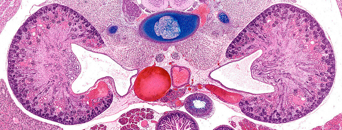
| Light micrograph of the developing kidneys of a foetus showing primitive renal corpuscles located around the outer cortex. Developing renal tubules and collecting ducts converge medially towards the large empty space termed the renal pelvis. In the postnatal kidney urine passes into the ureter via the pelvis. The aorta and inferior vena cava are seen in the midline, and a developing cartilaginous vertebral body and nucleus pulposus are stained blue. Paraffin section, alcian blue, and haematoxylin and eosin stain. Magnification: x16 when width printed at 10cm. | |
| Lizenzart: | Lizenzpflichtig |
| Credit: | Science Photo Library / Microscape |
| Bildgröße: | 7265 px × 2766 px |
| Modell-Rechte: | nicht erforderlich |
| Eigentums-Rechte: | nicht erforderlich |
| Restrictions: | - |
Preise für dieses Bild ab 15 €
Universitäten & Organisationen
(Informationsmaterial Digital, Informationsmaterial Print, Lehrmaterial Digital etc.)
ab 15 €
Redaktionell
(Bücher, Bücher: Sach- und Fachliteratur, Digitale Medien (redaktionell) etc.)
ab 30 €
Werbung
(Anzeigen, Aussenwerbung, Digitale Medien, Fernsehwerbung, Karten, Werbemittel, Zeitschriften etc.)
ab 55 €
Handelsprodukte
(bedruckte Textilie, Kalender, Postkarte, Grußkarte, Verpackung etc.)
ab 75 €
Pauschalpreise
Rechtepakete für die unbeschränkte Bildnutzung in Print oder Online
ab 495 €