Human seminiferous tubules, light micrographs
Bildnummer 13613992
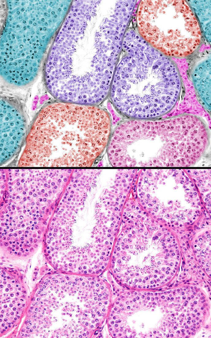
| Human testicle, light micrographs. The bottom micrograph shows seminiferous tubules. The development of spermatogenesis is asynchronous among seminiferous tubules and even within zones of a single seminiferous tubule. For this reason, in histological sections of the testis, seminiferous tubules can be seen in different phases of spermatogenesis. In the top micrograph, these different phases have been marked with different colours. Among the tubules are Leydig cells (pink). | |
| Lizenzart: | Lizenzpflichtig |
| Credit: | Science Photo Library / JOSE CALVO |
| Bildgröße: | 3840 px × 6144 px |
| Modell-Rechte: | nicht erforderlich |
| Eigentums-Rechte: | nicht erforderlich |
| Restrictions: | - |
Preise für dieses Bild ab 15 €
Universitäten & Organisationen
(Informationsmaterial Digital, Informationsmaterial Print, Lehrmaterial Digital etc.)
ab 15 €
Redaktionell
(Bücher, Bücher: Sach- und Fachliteratur, Digitale Medien (redaktionell) etc.)
ab 30 €
Werbung
(Anzeigen, Aussenwerbung, Digitale Medien, Fernsehwerbung, Karten, Werbemittel, Zeitschriften etc.)
ab 55 €
Handelsprodukte
(bedruckte Textilie, Kalender, Postkarte, Grußkarte, Verpackung etc.)
ab 75 €
Pauschalpreise
Rechtepakete für die unbeschränkte Bildnutzung in Print oder Online
ab 495 €