Brain haemorrhages, MRI angiogram
Bildnummer 13600174
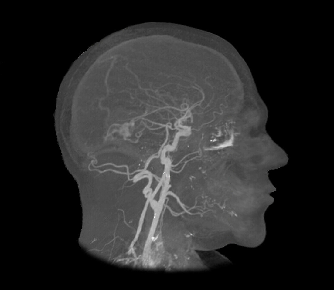
| Composite 2D and 3D magnetic resonance imaging (MRI) angiogram in right profile view of the brain of a 35-year-old patient hospitalized in the neurosurgical emergency room for attacks of severe headaches and suspected subarachnoid haemorrhage. An initial CT of the brain found a right parieto-occipital intra-parenchymal haemorrhage. This MRI reveals a subarachnoid haemorrhage secondary to a rupture of a right parieto-occipital arteriovenous malformation. | |
| Lizenzart: | Lizenzpflichtig |
| Credit: | Science Photo Library / Zephyr |
| Bildgröße: | 3969 px × 3450 px |
| Modell-Rechte: | nicht erforderlich |
| Eigentums-Rechte: | nicht erforderlich |
| Restrictions: | - |
Preise für dieses Bild ab 15 €
Universitäten & Organisationen
(Informationsmaterial Digital, Informationsmaterial Print, Lehrmaterial Digital etc.)
ab 15 €
Redaktionell
(Bücher, Bücher: Sach- und Fachliteratur, Digitale Medien (redaktionell) etc.)
ab 30 €
Werbung
(Anzeigen, Aussenwerbung, Digitale Medien, Fernsehwerbung, Karten, Werbemittel, Zeitschriften etc.)
ab 55 €
Handelsprodukte
(bedruckte Textilie, Kalender, Postkarte, Grußkarte, Verpackung etc.)
ab 75 €
Pauschalpreise
Rechtepakete für die unbeschränkte Bildnutzung in Print oder Online
ab 495 €