Arteriovenous malformation, composite MRI and 3D CT scan
Bildnummer 13599619
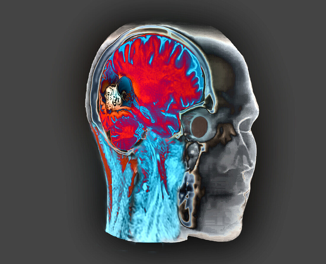
| Coloured composite magnetic resonance imaging (MRI) of a brain and 3D computed tomography (CT) scan of a 28-year-old female patient. The patient was admitted to emergency room with suspicion of subarachnoid haemorrhage (SAH) or arteriovenous malformation (AVM). The scan shows an arteriovenous malformation (purple) supplied by the distal branches of the right posterior cerebral artery (PCA) giving rise to a nidus (tangle of blood vessels) surrounded by a magma of draining veins flowing into the superior longitudinal sinus. | |
| Lizenzart: | Lizenzpflichtig |
| Credit: | Science Photo Library / Zephyr |
| Bildgröße: | 4194 px × 3402 px |
| Modell-Rechte: | nicht erforderlich |
| Eigentums-Rechte: | nicht erforderlich |
| Restrictions: | - |
Preise für dieses Bild ab 15 €
Universitäten & Organisationen
(Informationsmaterial Digital, Informationsmaterial Print, Lehrmaterial Digital etc.)
ab 15 €
Redaktionell
(Bücher, Bücher: Sach- und Fachliteratur, Digitale Medien (redaktionell) etc.)
ab 30 €
Werbung
(Anzeigen, Aussenwerbung, Digitale Medien, Fernsehwerbung, Karten, Werbemittel, Zeitschriften etc.)
ab 55 €
Handelsprodukte
(bedruckte Textilie, Kalender, Postkarte, Grußkarte, Verpackung etc.)
ab 75 €
Pauschalpreise
Rechtepakete für die unbeschränkte Bildnutzung in Print oder Online
ab 495 €