Traumatic brain injury, CT scan
Bildnummer 13599416
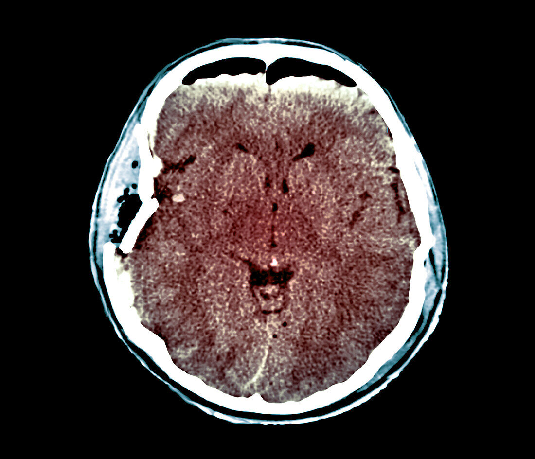
| Computed tomography (CT) scan in axial section of the brain of a 30 year old male patient with traumatic brain injury after a car accident. The CT shows a skull fracture of the right temporoparietal wall (left). There is a wound of the dura mater with presence of a frontal aeric bubble (top). Their is the wound with external and internal hemorrhagic contusion (brain bruises). The patient also has a diffuse internal cerebral oedema associated with a slight displacement of the ventricles. | |
| Lizenzart: | Lizenzpflichtig |
| Credit: | Science Photo Library / Zephyr |
| Bildgröße: | 3969 px × 3406 px |
| Modell-Rechte: | nicht erforderlich |
| Eigentums-Rechte: | nicht erforderlich |
| Restrictions: | - |
Preise für dieses Bild ab 15 €
Universitäten & Organisationen
(Informationsmaterial Digital, Informationsmaterial Print, Lehrmaterial Digital etc.)
ab 15 €
Redaktionell
(Bücher, Bücher: Sach- und Fachliteratur, Digitale Medien (redaktionell) etc.)
ab 30 €
Werbung
(Anzeigen, Aussenwerbung, Digitale Medien, Fernsehwerbung, Karten, Werbemittel, Zeitschriften etc.)
ab 55 €
Handelsprodukte
(bedruckte Textilie, Kalender, Postkarte, Grußkarte, Verpackung etc.)
ab 75 €
Pauschalpreise
Rechtepakete für die unbeschränkte Bildnutzung in Print oder Online
ab 495 €