Kidney cortex, light micrograph
Bildnummer 13586046
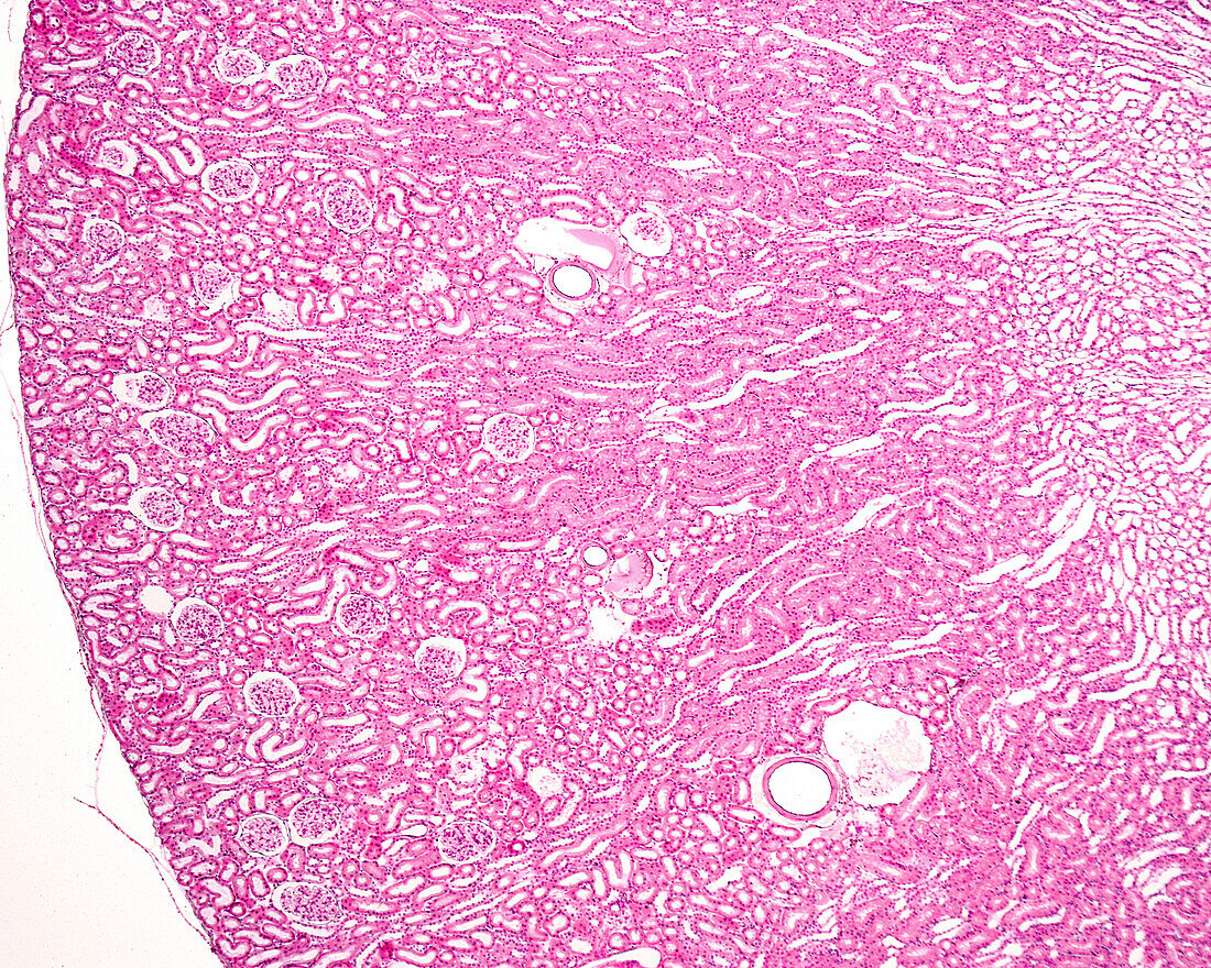
| Light micrograph of the renal cortex of an albino rat. The smaller size of the kidney makes it possible to observe the entire renal cortex and the border between the cortex and medulla in a single microscopic field. The tissue was fixed by perfusion, which explains the dilated appearance of the cortical arteries and veins. In the cortex, renal corpuscles are clearly visible. In the outer medulla (right), the difference between the outer stripe (similar to the cortex, but without renal corpuscles) and the inner stripe (which appears lighter, with cross-sectioned tubules) can be seen. | |
| Lizenzart: | Lizenzpflichtig |
| Credit: | Science Photo Library / JOSE CALVO |
| Bildgröße: | 3840 px × 3072 px |
| Modell-Rechte: | nicht erforderlich |
| Eigentums-Rechte: | nicht erforderlich |
| Restrictions: | - |
Preise für dieses Bild ab 15 €
Universitäten & Organisationen
(Informationsmaterial Digital, Informationsmaterial Print, Lehrmaterial Digital etc.)
ab 15 €
Redaktionell
(Bücher, Bücher: Sach- und Fachliteratur, Digitale Medien (redaktionell) etc.)
ab 30 €
Werbung
(Anzeigen, Aussenwerbung, Digitale Medien, Fernsehwerbung, Karten, Werbemittel, Zeitschriften etc.)
ab 55 €
Handelsprodukte
(bedruckte Textilie, Kalender, Postkarte, Grußkarte, Verpackung etc.)
ab 75 €
Pauschalpreise
Rechtepakete für die unbeschränkte Bildnutzung in Print oder Online
ab 495 €