Adrenal gland, light micrograph
Bildnummer 13585248
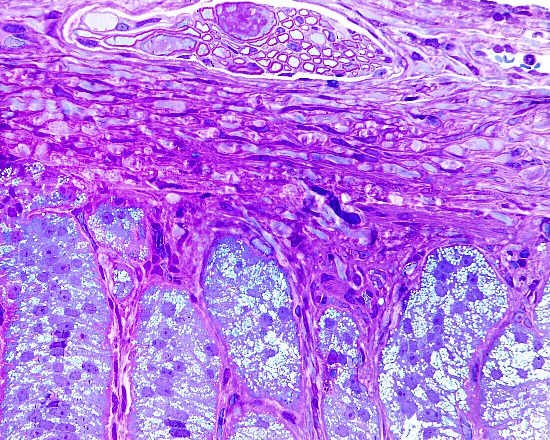
| Outer limit of the adrenal gland, light micrograph. The upper half of the image corresponds to the adrenal capsule, in which a small bundle of myelinated fibres can be seen, probably destined for the medulla. Below is the zona glomerulosa formed by elongated cells that are arranged in palisades perpendicular to the capsular surface, forming elongated ovoid nests. These cells have lipid droplets as is typical of steroid-secreting cells, although as can be seen here, the droplets are smaller and more numerous than in the zona fasciculata. 0.5 micrometre semi-fine cut of material embedded in plastic and stained with toluidine blue. | |
| Lizenzart: | Lizenzpflichtig |
| Credit: | Science Photo Library / JOSE CALVO |
| Bildgröße: | 3840 px × 3072 px |
| Modell-Rechte: | nicht erforderlich |
| Eigentums-Rechte: | nicht erforderlich |
| Restrictions: | - |
Preise für dieses Bild ab 15 €
Universitäten & Organisationen
(Informationsmaterial Digital, Informationsmaterial Print, Lehrmaterial Digital etc.)
ab 15 €
Redaktionell
(Bücher, Bücher: Sach- und Fachliteratur, Digitale Medien (redaktionell) etc.)
ab 30 €
Werbung
(Anzeigen, Aussenwerbung, Digitale Medien, Fernsehwerbung, Karten, Werbemittel, Zeitschriften etc.)
ab 55 €
Handelsprodukte
(bedruckte Textilie, Kalender, Postkarte, Grußkarte, Verpackung etc.)
ab 75 €
Pauschalpreise
Rechtepakete für die unbeschränkte Bildnutzung in Print oder Online
ab 495 €