Lung tissue damaged by mustard gas poisoning
Bildnummer 13525300
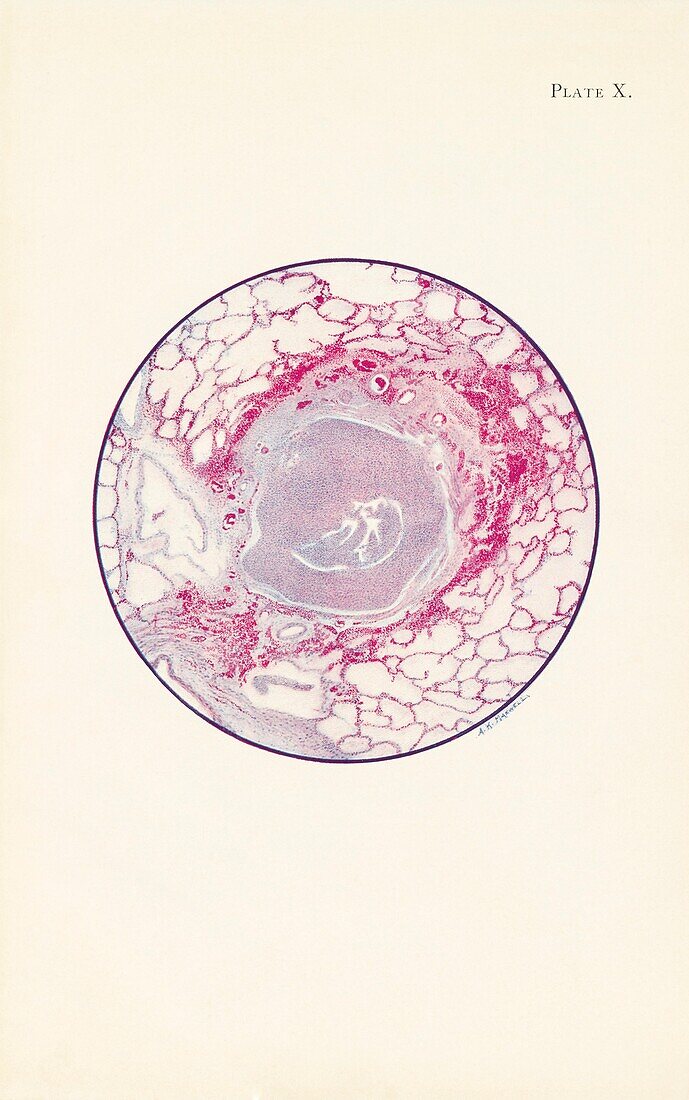
| Illustration showing a microscopic section of human lung tissue damaged by mustard gas poisoning during World War One. The bronchiole is filled with fibrin and pus cells. The lining of the epithelium has also been destroyed. A ring of haemorrhage is seen around the tissue surrounding the bronchiole. The patient from which the lung tissue was taken from, died 40 hours after being exposed to mustard gas. Mustard gas was used as a chemical warfare agent in the World War One. Exposure to mustard gas can cause coughing and shortness of breath in the short term. It also has long term effects such as mouth, throat and skin cancer as well as leukaemia. Illustration published in An Atlas of Gas Poisoning, 1918. | |
| Lizenzart: | Lizenzpflichtig |
| Credit: | Science Photo Library / Science History Institute |
| Bildgröße: | 3343 px × 5340 px |
| Modell-Rechte: | nicht erforderlich |
| Eigentums-Rechte: | nicht erforderlich |
| Restrictions: | - |
Preise für dieses Bild ab 15 €
Universitäten & Organisationen
(Informationsmaterial Digital, Informationsmaterial Print, Lehrmaterial Digital etc.)
ab 15 €
Redaktionell
(Bücher, Bücher: Sach- und Fachliteratur, Digitale Medien (redaktionell) etc.)
ab 30 €
Werbung
(Anzeigen, Aussenwerbung, Digitale Medien, Fernsehwerbung, Karten, Werbemittel, Zeitschriften etc.)
ab 55 €
Handelsprodukte
(bedruckte Textilie, Kalender, Postkarte, Grußkarte, Verpackung etc.)
ab 75 €
Pauschalpreise
Rechtepakete für die unbeschränkte Bildnutzung in Print oder Online
ab 495 €