Thyroid gland follicles, light micrograph
Bildnummer 13511482
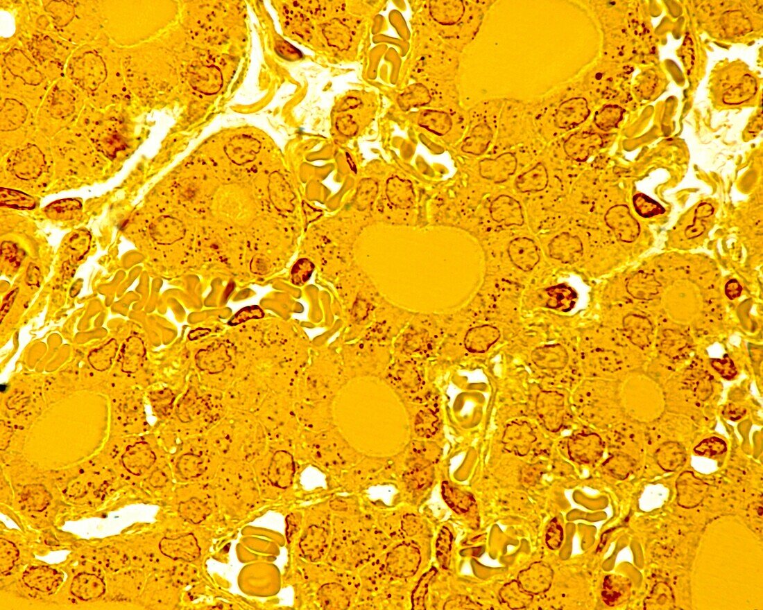
| Light micrograph of thyroid follicles with a homogeneous central colloid, filling the entire follicular cavity, surrounded by a cubic to prismatic epithelium. Unlike routine histologic techniques where artifacts in the colloid are the rule, the use of more refined fixation and embedding in this material provides a perfect image of the structure of the thyroid follicles. The dark brown granulations present in the cytoplasm of thyrocytes are lysosomes. Surrounding the follicles are abundant blood capillaries. 0.5 micrometre thick semithin section of plastic-embedded material stained with silver method. | |
| Lizenzart: | Lizenzpflichtig |
| Credit: | Science Photo Library / JOSE CALVO |
| Bildgröße: | 3840 px × 3072 px |
| Modell-Rechte: | nicht erforderlich |
| Eigentums-Rechte: | nicht erforderlich |
| Restrictions: | - |
Preise für dieses Bild ab 15 €
Universitäten & Organisationen
(Informationsmaterial Digital, Informationsmaterial Print, Lehrmaterial Digital etc.)
ab 15 €
Redaktionell
(Bücher, Bücher: Sach- und Fachliteratur, Digitale Medien (redaktionell) etc.)
ab 30 €
Werbung
(Anzeigen, Aussenwerbung, Digitale Medien, Fernsehwerbung, Karten, Werbemittel, Zeitschriften etc.)
ab 55 €
Handelsprodukte
(bedruckte Textilie, Kalender, Postkarte, Grußkarte, Verpackung etc.)
ab 75 €
Pauschalpreise
Rechtepakete für die unbeschränkte Bildnutzung in Print oder Online
ab 495 €
Keywords
- Anatomie,
- anatomisch,
- Biologie,
- biologisch,
- Drüse,
- endokrin,
- Endokrinologie,
- Endokrinsystem,
- Follikel,
- gesund,
- Histologie,
- histologisch,
- Hormone,
- hormonell,
- Lichtmikroskop,
- lichtmikroskopische Aufnahme,
- Mensch,
- menschlicher Körper,
- Mikroskopie,
- Niemand,
- normal,
- Schilddrüse,
- Schilddrüsenfollikel,
- Thyreoidea,
- Zelle