Parathyroid gland, light micrograph
Bildnummer 13506087
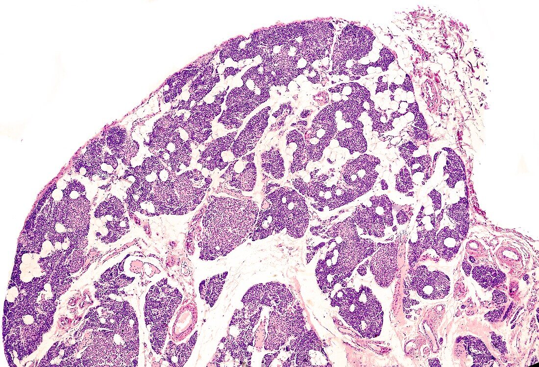
| Very low magnification light micrograph showing the parathyroid gland of a young subject. At this time, most of the volume of the gland corresponds to parenchyma. Adipose tissue is beginning to appear in the connective tissue septa, although it is not yet abundant. The parenchyma is formed by thick cellular cords integrated, for the most part, by very small main cells, with nuclei very close to each other. Oxyphilic cells already exist that, at some points, form nodules of considerable size. | |
| Lizenzart: | Lizenzpflichtig |
| Credit: | Science Photo Library / JOSE CALVO |
| Bildgröße: | 3840 px × 2613 px |
| Modell-Rechte: | nicht erforderlich |
| Eigentums-Rechte: | nicht erforderlich |
| Restrictions: | - |
Preise für dieses Bild ab 15 €
Universitäten & Organisationen
(Informationsmaterial Digital, Informationsmaterial Print, Lehrmaterial Digital etc.)
ab 15 €
Redaktionell
(Bücher, Bücher: Sach- und Fachliteratur, Digitale Medien (redaktionell) etc.)
ab 30 €
Werbung
(Anzeigen, Aussenwerbung, Digitale Medien, Fernsehwerbung, Karten, Werbemittel, Zeitschriften etc.)
ab 55 €
Handelsprodukte
(bedruckte Textilie, Kalender, Postkarte, Grußkarte, Verpackung etc.)
ab 75 €
Pauschalpreise
Rechtepakete für die unbeschränkte Bildnutzung in Print oder Online
ab 495 €