Spinal sensory ganglion, light micrograph
Bildnummer 13505124
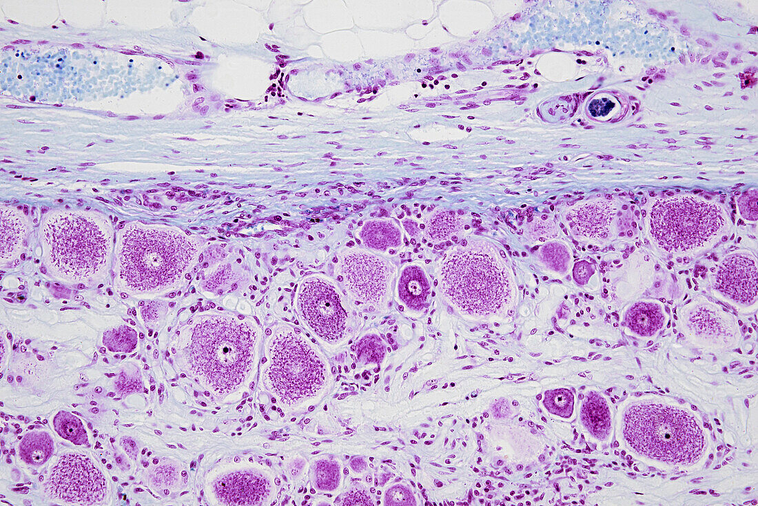
| Light micrograph of a spinal sensory ganglion, which is also known as a dorsal root ganglion. The ganglion is formed of a cluster of nerve cell bodies each with a central nucleus and densely stained cytoplasm (purple). They are surrounded by many small satellite or glial cells. Sensory signals from peripheral organs travel via nerves towards the spinal cord and pass through the ganglion before entering the grey matter (dorsal horn) of the spinal cord. At top is the connective tissue (light blue) capsule that surrounds the ganglion. Within it are blood vessels carrying red blood cells (darker blue). Magnification x200 when printed at 15cm wide. | |
| Lizenzart: | Lizenzpflichtig |
| Credit: | Science Photo Library / EYE OF SCIENCE |
| Bildgröße: | 6016 px × 4016 px |
| Modell-Rechte: | nicht erforderlich |
| Eigentums-Rechte: | nicht erforderlich |
| Restrictions: |
|
Preise für dieses Bild ab 15 €
Universitäten & Organisationen
(Informationsmaterial Digital, Informationsmaterial Print, Lehrmaterial Digital etc.)
ab 15 €
Redaktionell
(Bücher, Bücher: Sach- und Fachliteratur, Digitale Medien (redaktionell) etc.)
ab 30 €
Werbung
(Anzeigen, Aussenwerbung, Digitale Medien, Fernsehwerbung, Karten, Werbemittel, Zeitschriften etc.)
ab 55 €
Handelsprodukte
(bedruckte Textilie, Kalender, Postkarte, Grußkarte, Verpackung etc.)
ab 75 €
Pauschalpreise
Rechtepakete für die unbeschränkte Bildnutzung in Print oder Online
ab 495 €