Urinary bladder wall, light micrograph
Bildnummer 13504657
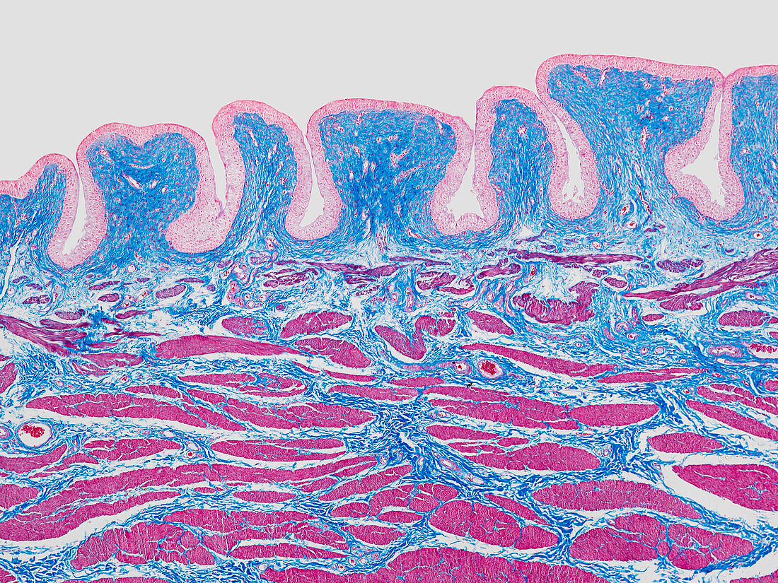
| Light micrograph of a section through the wall of the urinary bladder in its contracted (empty) state. The numerous folds, or rugae, allow the bladder to expand as it fills. Lining the interior of the bladder is transitional epithelium (pink), which is able to expand, but also forms an impermeable barrier to prevent toxins from the urine entering the blood stream. Beneath the epithelium is connective tissue (blue) with blood vessels and smooth muscle cells (red, across bottom). Magnification: x40 when printed at 15cm wide. | |
| Lizenzart: | Lizenzpflichtig |
| Credit: | Science Photo Library / EYE OF SCIENCE |
| Bildgröße: | 5354 px × 4016 px |
| Modell-Rechte: | nicht erforderlich |
| Eigentums-Rechte: | nicht erforderlich |
| Restrictions: |
|
Preise für dieses Bild ab 15 €
Universitäten & Organisationen
(Informationsmaterial Digital, Informationsmaterial Print, Lehrmaterial Digital etc.)
ab 15 €
Redaktionell
(Bücher, Bücher: Sach- und Fachliteratur, Digitale Medien (redaktionell) etc.)
ab 30 €
Werbung
(Anzeigen, Aussenwerbung, Digitale Medien, Fernsehwerbung, Karten, Werbemittel, Zeitschriften etc.)
ab 55 €
Handelsprodukte
(bedruckte Textilie, Kalender, Postkarte, Grußkarte, Verpackung etc.)
ab 75 €
Pauschalpreise
Rechtepakete für die unbeschränkte Bildnutzung in Print oder Online
ab 495 €