Foetal finger joint, light micrograph
Bildnummer 13504518
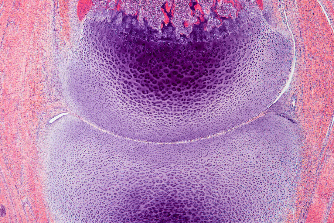
| Light micrograph of a longitudinal section through a foetal finger joint. The finger bones are initially formed of cartilage (purple). Bone formation (ossification) starts from the middle of the cartilage and proceeds outwards. Areas where bone has already formed are red, areas of active ossification are dark purple. Either side of the bones are tendons and muscles. Magnification: x40 when printed at 15cm wide. | |
| Lizenzart: | Lizenzpflichtig |
| Credit: | Science Photo Library / EYE OF SCIENCE |
| Bildgröße: | 6016 px × 4016 px |
| Modell-Rechte: | nicht erforderlich |
| Eigentums-Rechte: | nicht erforderlich |
| Restrictions: |
|
Preise für dieses Bild ab 15 €
Universitäten & Organisationen
(Informationsmaterial Digital, Informationsmaterial Print, Lehrmaterial Digital etc.)
ab 15 €
Redaktionell
(Bücher, Bücher: Sach- und Fachliteratur, Digitale Medien (redaktionell) etc.)
ab 30 €
Werbung
(Anzeigen, Aussenwerbung, Digitale Medien, Fernsehwerbung, Karten, Werbemittel, Zeitschriften etc.)
ab 55 €
Handelsprodukte
(bedruckte Textilie, Kalender, Postkarte, Grußkarte, Verpackung etc.)
ab 75 €
Pauschalpreise
Rechtepakete für die unbeschränkte Bildnutzung in Print oder Online
ab 495 €