Small intestine, light micrograph
Bildnummer 13504491
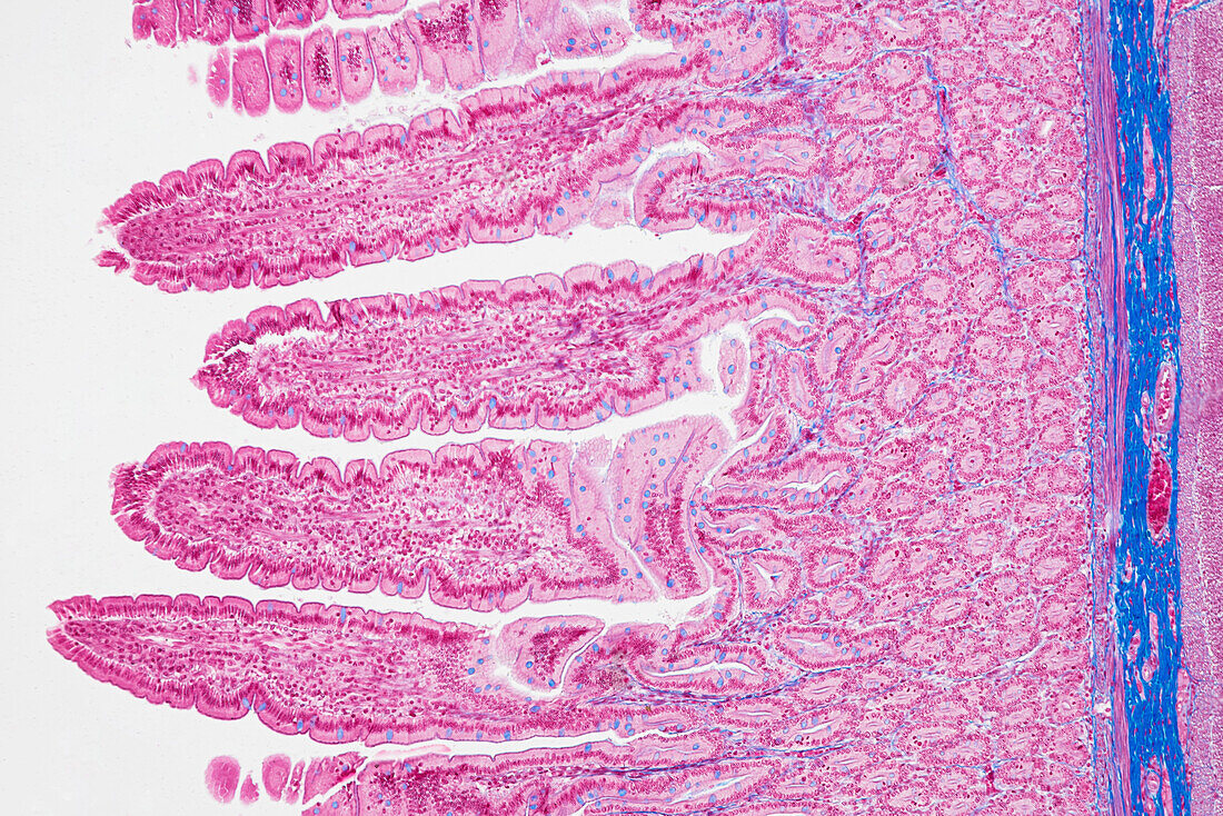
| Light micrograph of a section through a duodenum, the beginning of the small intestine, which runs from the stomach to the large intestine. It is where digestion begins and nutrients are absorbed into the blood. The interior (lumen, left) is lined with villi, which are folds in the intestinal surface that greatly increase the surface area for absorption. Also within the surface are crypts of Lieberkuhn (red-lined ovoids), which secrete enzymes into the lumen that help to digest the food. Underlying this layer is the muscularis mucosae (thin violet line) and connective tissue (blue). Across bottom are layers of smooth muscle and then circular and longitudinal muscles that contract and relax to move food through the duodenum. Magnification: x100 when printed at 15 centimetres wide. | |
| Lizenzart: | Lizenzpflichtig |
| Credit: | Science Photo Library / EYE OF SCIENCE |
| Bildgröße: | 6016 px × 4016 px |
| Modell-Rechte: | nicht erforderlich |
| Eigentums-Rechte: | nicht erforderlich |
| Restrictions: |
|
Preise für dieses Bild ab 15 €
Universitäten & Organisationen
(Informationsmaterial Digital, Informationsmaterial Print, Lehrmaterial Digital etc.)
ab 15 €
Redaktionell
(Bücher, Bücher: Sach- und Fachliteratur, Digitale Medien (redaktionell) etc.)
ab 30 €
Werbung
(Anzeigen, Aussenwerbung, Digitale Medien, Fernsehwerbung, Karten, Werbemittel, Zeitschriften etc.)
ab 55 €
Handelsprodukte
(bedruckte Textilie, Kalender, Postkarte, Grußkarte, Verpackung etc.)
ab 75 €
Pauschalpreise
Rechtepakete für die unbeschränkte Bildnutzung in Print oder Online
ab 495 €
Keywords
- Anatomie,
- anatomisch,
- Bindegewebe,
- Biologie,
- biologisch,
- Darm,
- Darm-,
- Dünndarm,
- gastrointestinal,
- Gedärme,
- gesund,
- GI tract,
- Histologie,
- histologisch,
- Hund,
- Krypten von Lieberkuhn,
- lichtmikroskopische Aufnahme,
- Lumen,
- Mikroskopie,
- Muscularis mucosae,
- Muskel,
- Niemand,
- normal,
- Säugetier,
- Säugetier-,
- Sektion,
- sektioniert,
- Verdauung,
- Verdauungskanal,
- Verdauungssystem,
- Zelle,
- Zellen,
- Zotte,
- Zotten