Colour Enhanced Primitive Neuroectodermal Tumour (PNET) MRI
Bildnummer 13496238
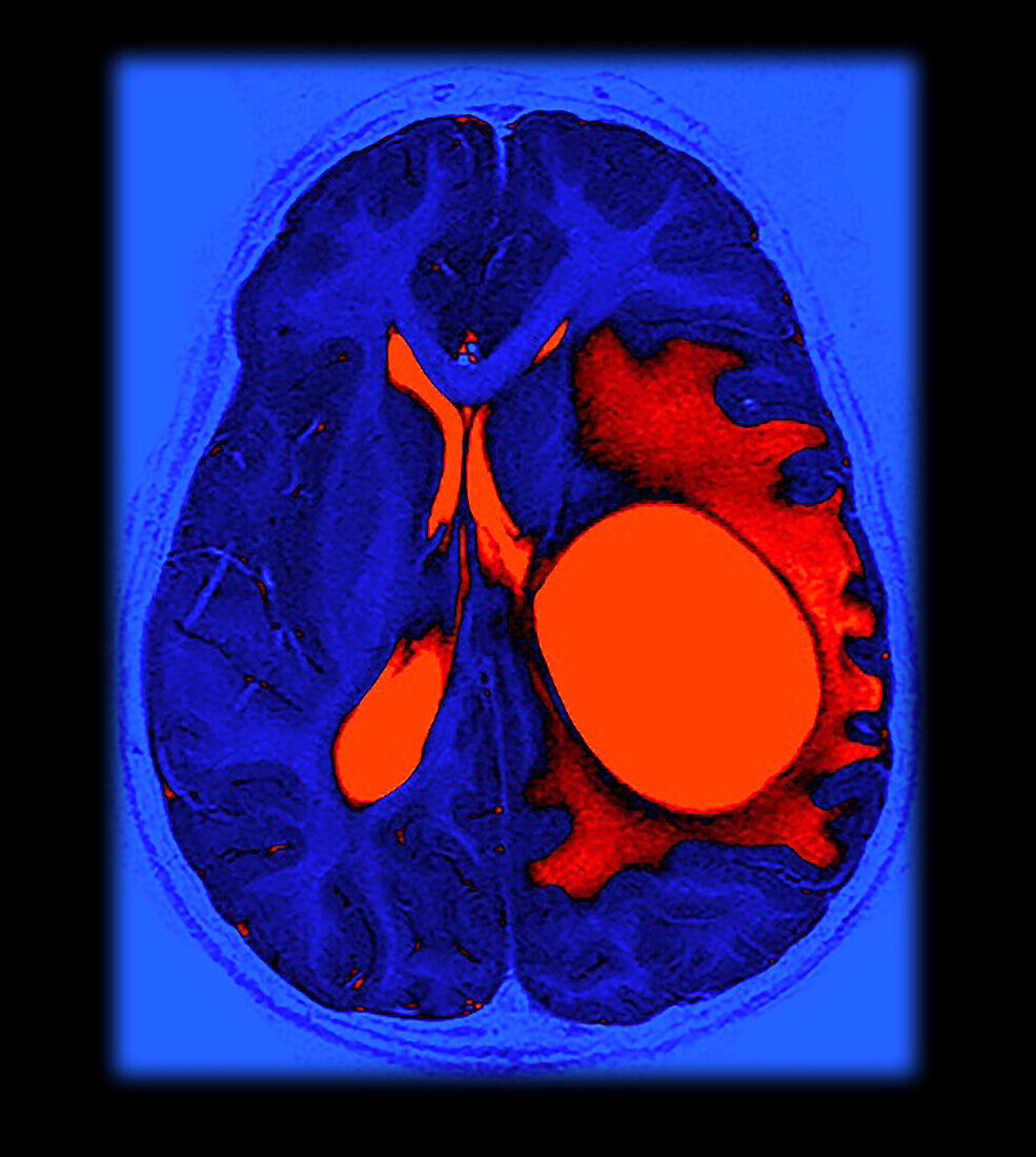
| This colour enhanced axial (cross sectional) T2 weighted MR image shows a large cystic and partially solid mass in the fronto-parietal region representing a malignant brain tumour in an infant called a PNET (primitive neuroectodermal tumour). | |
| Lizenzart: | Lizenzpflichtig |
| Credit: | Science Photo Library / Science Source / Living Art Enterprises, LLC |
| Bildgröße: | 3900 px × 4356 px |
| Modell-Rechte: | nicht erforderlich |
| Eigentums-Rechte: | nicht erforderlich |
| Restrictions: | - |
Preise für dieses Bild ab 15 €
Universitäten & Organisationen
(Informationsmaterial Digital, Informationsmaterial Print, Lehrmaterial Digital etc.)
ab 15 €
Redaktionell
(Bücher, Bücher: Sach- und Fachliteratur, Digitale Medien (redaktionell) etc.)
ab 30 €
Werbung
(Anzeigen, Aussenwerbung, Digitale Medien, Fernsehwerbung, Karten, Werbemittel, Zeitschriften etc.)
ab 55 €
Handelsprodukte
(bedruckte Textilie, Kalender, Postkarte, Grußkarte, Verpackung etc.)
ab 75 €
Pauschalpreise
Rechtepakete für die unbeschränkte Bildnutzung in Print oder Online
ab 495 €