Aqueductal Stenosis, Hydrocephalus, MRI
Bildnummer 13496131
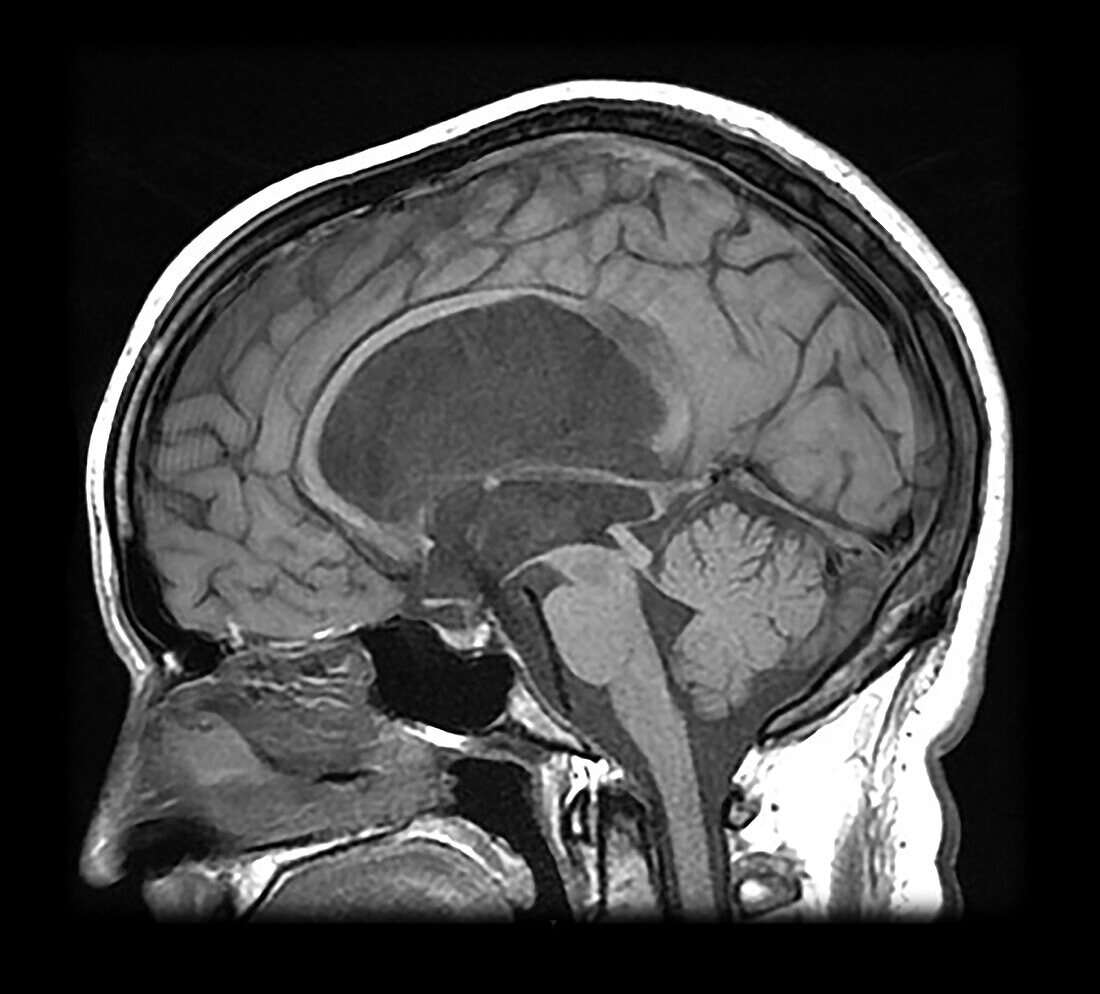
| This sagittal T1 weighted MR image shows dilated third and lateral ventricles with a normal size fourth ventricle. Stenosis with occlusion of the lower aqueduct of Sylvius is present. Surgical intervention has been performed with an inferior third ventriculostomy which is patent as demonstrated by a prominent flow void of CSF flow from the third ventricle to the pre-pontine cistern. | |
| Lizenzart: | Lizenzpflichtig |
| Credit: | Science Photo Library / Science Source / Living Art Enterprises, LLC |
| Bildgröße: | 4318 px × 3900 px |
| Modell-Rechte: | nicht erforderlich |
| Eigentums-Rechte: | nicht erforderlich |
| Restrictions: | - |
Preise für dieses Bild ab 15 €
Universitäten & Organisationen
(Informationsmaterial Digital, Informationsmaterial Print, Lehrmaterial Digital etc.)
ab 15 €
Redaktionell
(Bücher, Bücher: Sach- und Fachliteratur, Digitale Medien (redaktionell) etc.)
ab 30 €
Werbung
(Anzeigen, Aussenwerbung, Digitale Medien, Fernsehwerbung, Karten, Werbemittel, Zeitschriften etc.)
ab 55 €
Handelsprodukte
(bedruckte Textilie, Kalender, Postkarte, Grußkarte, Verpackung etc.)
ab 75 €
Pauschalpreise
Rechtepakete für die unbeschränkte Bildnutzung in Print oder Online
ab 495 €