Reticular fibres in human pituitary gland, light micrograph
Bildnummer 13478039
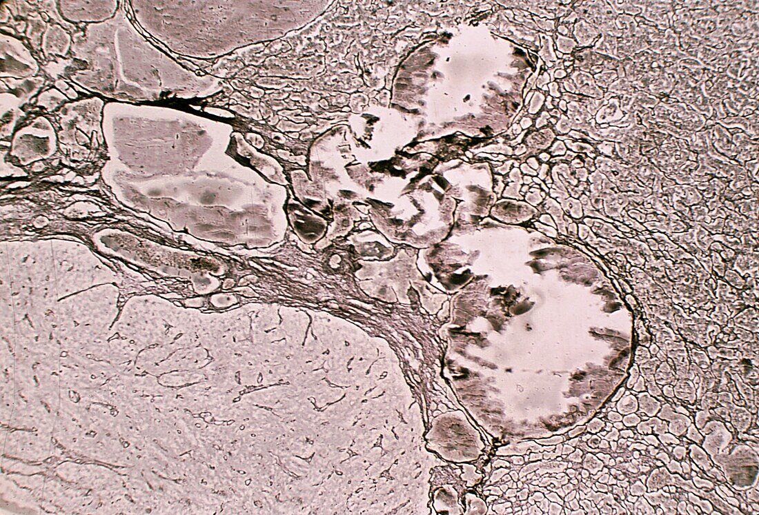
| Reticular fibres in human pituitary gland, light micrograph. This is the border between the anterior lobe (right), intermediate lobe (centre) and posterior lobe (left) of the pituitary. It has been stained with the silver method to highlight reticular fibres. With this method, the differences in the organization pattern of each of these lobes stand out. The anterior lobe shows a cell cords pattern. The intermediate lobe contains large vesicles and cysts (whose content appears fragmented by cutting artifacts). The posterior lobe has poor reticulin development, limited to the wall of the blood vessels. | |
| Lizenzart: | Lizenzpflichtig |
| Credit: | Science Photo Library / JOSE CALVO |
| Bildgröße: | 3840 px × 2609 px |
| Modell-Rechte: | nicht erforderlich |
| Eigentums-Rechte: | nicht erforderlich |
| Restrictions: | - |
Preise für dieses Bild ab 15 €
Universitäten & Organisationen
(Informationsmaterial Digital, Informationsmaterial Print, Lehrmaterial Digital etc.)
ab 15 €
Redaktionell
(Bücher, Bücher: Sach- und Fachliteratur, Digitale Medien (redaktionell) etc.)
ab 30 €
Werbung
(Anzeigen, Aussenwerbung, Digitale Medien, Fernsehwerbung, Karten, Werbemittel, Zeitschriften etc.)
ab 55 €
Handelsprodukte
(bedruckte Textilie, Kalender, Postkarte, Grußkarte, Verpackung etc.)
ab 75 €
Pauschalpreise
Rechtepakete für die unbeschränkte Bildnutzung in Print oder Online
ab 495 €