Aortic aneurysm, CT scan
Bildnummer 13465296
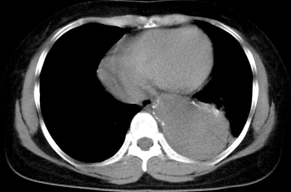
| Contrast chest computed tomography (CT) scan of 54-year-old female patient showing enlargement of the descending thoracic aorta. There is also calcification of the aortic wall. The lumen of the aorta is hyperdense (bright). The hypodense (slightly dark) area is a thrombus (blood clot) in the artery. | |
| Lizenzart: | Lizenzfrei |
| Credit: | Science Photo Library / RAJAAISYA |
| Modell-Rechte: | nicht erforderlich |
| Eigentums-Rechte: | nicht erforderlich |
| Restrictions: | - |
Preise für dieses Bild ab 29 €
Für digitale Nutzung (72 dpi)
ab 29 €
Für Druckauflösung (300 dpi)
ab 300 €