Second degree type II atrioventricular block, illustration
Bildnummer 13453435
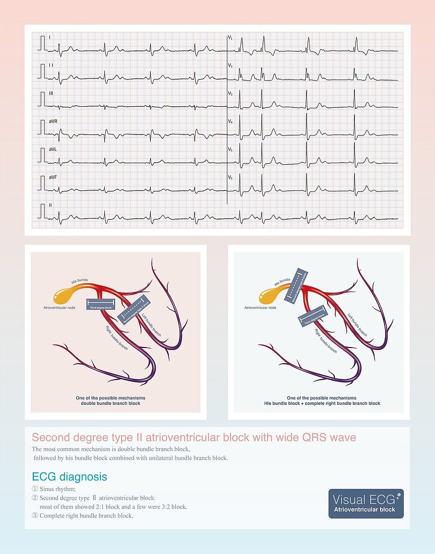
| Electrocardiogram (ECG) illustration from a 38 year old female who was admitted to hospital with repeated syncope for 3 years. The electrocardiogram showed sinus rhythm, second degree type II atrioventricular block, and complete right bundle branch block. From the electrocardiogram it was deduced that the patient has a high possibility of double bundle branch block, and finally a permanent artificial cardiac pacemaker was inserted. | |
| Lizenzart: | Lizenzpflichtig |
| Credit: | Science Photo Library / CHONGQING TUMI TECHNOLOGY LTD |
| Bildgröße: | 3200 px × 4077 px |
| Modell-Rechte: | nicht erforderlich |
| Eigentums-Rechte: | nicht erforderlich |
| Restrictions: | - |
Preise für dieses Bild ab 15 €
Universitäten & Organisationen
(Informationsmaterial Digital, Informationsmaterial Print, Lehrmaterial Digital etc.)
ab 15 €
Redaktionell
(Bücher, Bücher: Sach- und Fachliteratur, Digitale Medien (redaktionell) etc.)
ab 30 €
Werbung
(Anzeigen, Aussenwerbung, Digitale Medien, Fernsehwerbung, Karten, Werbemittel, Zeitschriften etc.)
ab 55 €
Handelsprodukte
(bedruckte Textilie, Kalender, Postkarte, Grußkarte, Verpackung etc.)
ab 75 €
Pauschalpreise
Rechtepakete für die unbeschränkte Bildnutzung in Print oder Online
ab 495 €