Rheumatoid arthritis, X-ray
Bildnummer 13453110
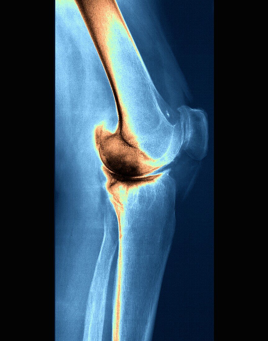
| X-ray image of the left knee of a 54-year-old female patient with rheumatoid arthritis. Rheumatoid arthritis is an autoimmune disease that attacks the joints, leading to painful, stiff and deformed joints. The scan shows diffuse bone demineralization producing a heterogeneous appearance by the existence of geodic images of internal femoral condylar and external and especially the internal tibial plateau. The patient also has bicompartmental knee osteoarthritis (osteoarthritis affecting two of three compartments of the knee joint). | |
| Lizenzart: | Lizenzpflichtig |
| Credit: | Science Photo Library / Zephyr |
| Bildgröße: | 3402 px × 4336 px |
| Modell-Rechte: | nicht erforderlich |
| Eigentums-Rechte: | nicht erforderlich |
| Restrictions: | - |
Preise für dieses Bild ab 15 €
Universitäten & Organisationen
(Informationsmaterial Digital, Informationsmaterial Print, Lehrmaterial Digital etc.)
ab 15 €
Redaktionell
(Bücher, Bücher: Sach- und Fachliteratur, Digitale Medien (redaktionell) etc.)
ab 30 €
Werbung
(Anzeigen, Aussenwerbung, Digitale Medien, Fernsehwerbung, Karten, Werbemittel, Zeitschriften etc.)
ab 55 €
Handelsprodukte
(bedruckte Textilie, Kalender, Postkarte, Grußkarte, Verpackung etc.)
ab 75 €
Pauschalpreise
Rechtepakete für die unbeschränkte Bildnutzung in Print oder Online
ab 495 €