Bicornuate uterus, X-ray
Bildnummer 13452635
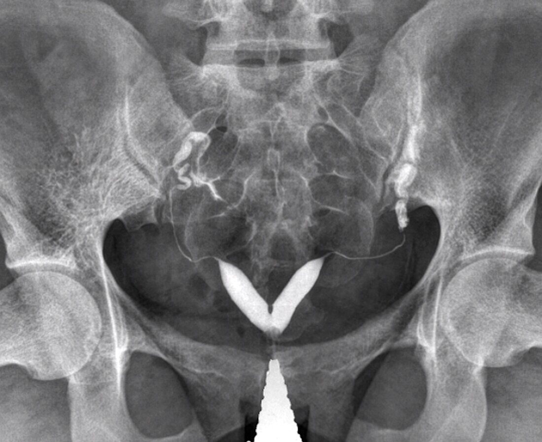
| X-ray of the abdomen of a 24 year old female patient with a two-horned (bicornuate) uterus (white, lower centre). This congenital defect results in the division of the upper end of the uterus into two halves. It may result in a higher rate of miscarriage during pregnancy, or promote premature labour or a breech (buttocks first) presentation at birth. Surgical treatments for a bicornuate uterus include reshaping the uterus to create a larger uterine cavity, and stitching shut the neck of the uterus (cervix, bottom centre) during pregnancy to prevent its early opening. A contrast medium, introduced through the cervix, reveals the position of the uterus on the X-ray. | |
| Lizenzart: | Lizenzpflichtig |
| Credit: | Science Photo Library / Zephyr |
| Bildgröße: | 4102 px × 3354 px |
| Modell-Rechte: | nicht erforderlich |
| Eigentums-Rechte: | nicht erforderlich |
| Restrictions: | - |
Preise für dieses Bild ab 15 €
Universitäten & Organisationen
(Informationsmaterial Digital, Informationsmaterial Print, Lehrmaterial Digital etc.)
ab 15 €
Redaktionell
(Bücher, Bücher: Sach- und Fachliteratur, Digitale Medien (redaktionell) etc.)
ab 30 €
Werbung
(Anzeigen, Aussenwerbung, Digitale Medien, Fernsehwerbung, Karten, Werbemittel, Zeitschriften etc.)
ab 55 €
Handelsprodukte
(bedruckte Textilie, Kalender, Postkarte, Grußkarte, Verpackung etc.)
ab 75 €
Pauschalpreise
Rechtepakete für die unbeschränkte Bildnutzung in Print oder Online
ab 495 €
Keywords
- abnormal,
- angeboren,
- Anomalie,
- Ausrüstung,
- defekt,
- Deformiert,
- Deformität,
- Diagnose,
- Einfarbig,
- Fortpflanzungssystem,
- Frau,
- Gebärmutter,
- Geburt,
- Geburtsfehler,
- geduldig,
- Gesundheitswesen,
- Gynäkologie,
- Horn,
- Hörner,
- Kondition,
- Kontrastmittel,
- Medizin,
- medizinisch,
- menschlicher Körper,
- Niemand,
- Radiographie,
- Reproduktionspathologie,
- Röntgen,
- Röntgenbild,
- Salpingogramm,
- schwarz und weiß,
- Störung,
- ungesund,
- uterin,
- Uterus,
- Weiblich,
- weibliches Fortpflanzungssystem