Synovial membrane, light micrograph
Bildnummer 13452259
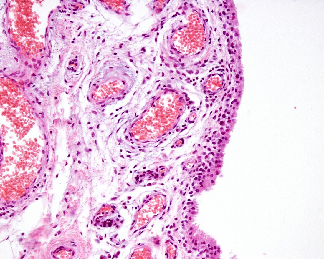
| Light micrograph of a human synovial membrane (innermost part of the joint capsule). It delimits the joint cavity of the diarthrosis. In the vicinity of the lumen of the joint cavity, a band of synovial cells is concentrated, adopting an appearance reminiscent of a lining epithelium, however, these synovial cells are conjunctive tissue cells that form a discontinuous lining. At left is a space occupied by loose connective tissue with dilated blood vessels with abundant red cells in the lumen, indicating congestion. | |
| Lizenzart: | Lizenzpflichtig |
| Credit: | Science Photo Library / JOSE CALVO |
| Bildgröße: | 3840 px × 3072 px |
| Modell-Rechte: | nicht erforderlich |
| Eigentums-Rechte: | nicht erforderlich |
| Restrictions: | - |
Preise für dieses Bild ab 15 €
Universitäten & Organisationen
(Informationsmaterial Digital, Informationsmaterial Print, Lehrmaterial Digital etc.)
ab 15 €
Redaktionell
(Bücher, Bücher: Sach- und Fachliteratur, Digitale Medien (redaktionell) etc.)
ab 30 €
Werbung
(Anzeigen, Aussenwerbung, Digitale Medien, Fernsehwerbung, Karten, Werbemittel, Zeitschriften etc.)
ab 55 €
Handelsprodukte
(bedruckte Textilie, Kalender, Postkarte, Grußkarte, Verpackung etc.)
ab 75 €
Pauschalpreise
Rechtepakete für die unbeschränkte Bildnutzung in Print oder Online
ab 495 €