Tendon, light micrograph
Bildnummer 13452243
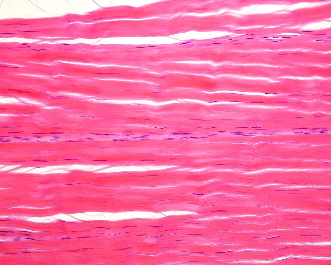
| Light micrograph of a longitudinal section of a tendon. The tendon is made up of a connective tissue very rich in collagen fibres that are arranged in bundles(red bands stained with eosin). Few narrow and elongated nuclei are seen between these bundles, corresponding to a particular type of fibroblasts called tenocytes. At centre there is a band of nuclei that corresponds to a connective tissue sheath that encompasses the bundles of collagen bundles and is called the internal peritenon. | |
| Lizenzart: | Lizenzpflichtig |
| Credit: | Science Photo Library / JOSE CALVO |
| Bildgröße: | 3840 px × 3072 px |
| Modell-Rechte: | nicht erforderlich |
| Eigentums-Rechte: | nicht erforderlich |
| Restrictions: | - |
Preise für dieses Bild ab 15 €
Universitäten & Organisationen
(Informationsmaterial Digital, Informationsmaterial Print, Lehrmaterial Digital etc.)
ab 15 €
Redaktionell
(Bücher, Bücher: Sach- und Fachliteratur, Digitale Medien (redaktionell) etc.)
ab 30 €
Werbung
(Anzeigen, Aussenwerbung, Digitale Medien, Fernsehwerbung, Karten, Werbemittel, Zeitschriften etc.)
ab 55 €
Handelsprodukte
(bedruckte Textilie, Kalender, Postkarte, Grußkarte, Verpackung etc.)
ab 75 €
Pauschalpreise
Rechtepakete für die unbeschränkte Bildnutzung in Print oder Online
ab 495 €