Compact bone osteons, polarised light micrograph
Bildnummer 13452242
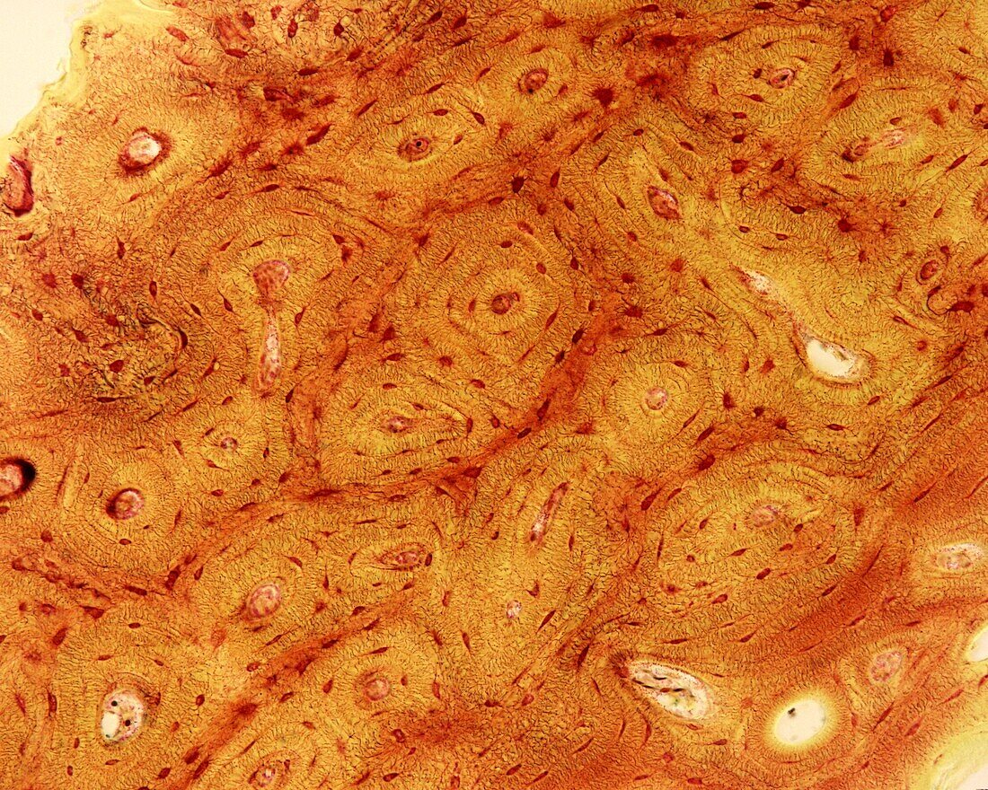
| Light micrograph of compact bone belonging to a cross-sectioned long bone diaphysis, stained with the Schmorl technique. A variant of this technique has been used that stains the somas and fine processes of osteocytes reddish. Osteons are clearly identified, being the essential components of compact bone tissue. They correspond to rounded or oval structures, made up of bony lamellae arranged concentrically around a small duct called (Haversian duct), occupied by a small blood vessel. Between the lamellae are the oval bodies of the osteocytes, from which numerous and fine extensions come into contact with each other, forming a complex labyrinth. Among the osteons there are groupings of bone lamellae called interstitial lamellae. | |
| Lizenzart: | Lizenzpflichtig |
| Credit: | Science Photo Library / JOSE CALVO |
| Bildgröße: | 3840 px × 3072 px |
| Modell-Rechte: | nicht erforderlich |
| Eigentums-Rechte: | nicht erforderlich |
| Restrictions: | - |
Preise für dieses Bild ab 15 €
Universitäten & Organisationen
(Informationsmaterial Digital, Informationsmaterial Print, Lehrmaterial Digital etc.)
ab 15 €
Redaktionell
(Bücher, Bücher: Sach- und Fachliteratur, Digitale Medien (redaktionell) etc.)
ab 30 €
Werbung
(Anzeigen, Aussenwerbung, Digitale Medien, Fernsehwerbung, Karten, Werbemittel, Zeitschriften etc.)
ab 55 €
Handelsprodukte
(bedruckte Textilie, Kalender, Postkarte, Grußkarte, Verpackung etc.)
ab 75 €
Pauschalpreise
Rechtepakete für die unbeschränkte Bildnutzung in Print oder Online
ab 495 €