Embryonic bone diaphysis, light micrograph
Bildnummer 13452216
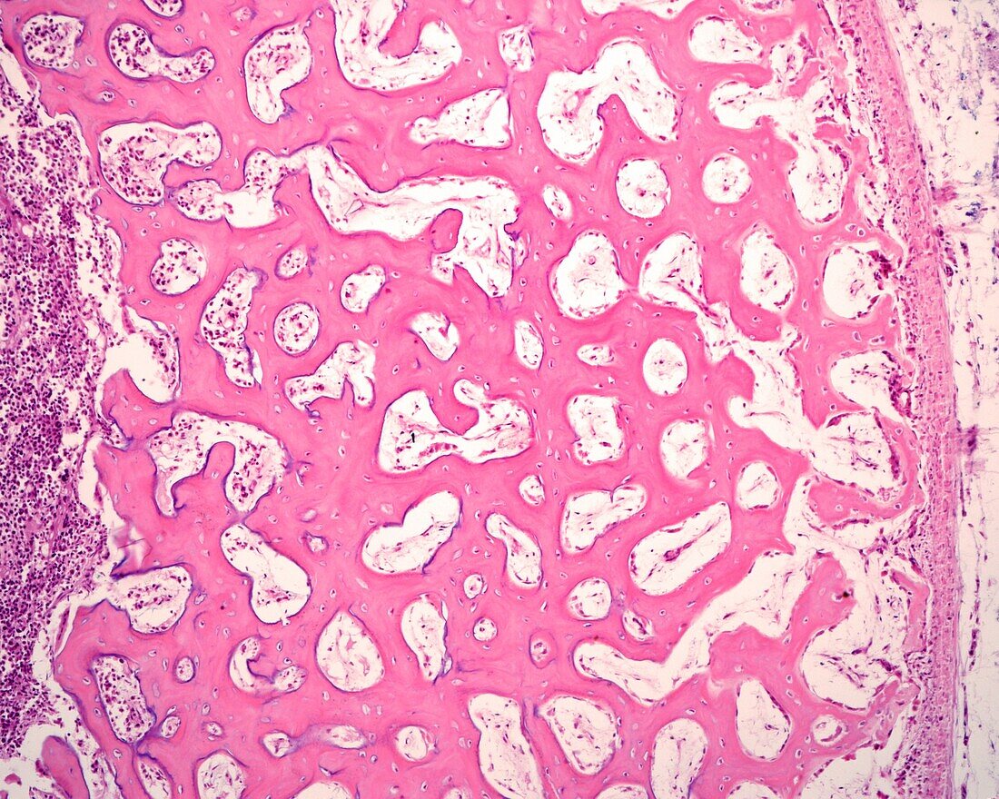
| Peripheral area of the diaphysis of an embryonic bone, light micrograph. The periosteum is at right, showing two layers: an outer, or fibrous layer (pink), and an internal, or osteogenic layer, with a more cellular mesenchyme, where the osteoprogenitor cells are generated. In the more peripheral bone trabeculae, osteoid lines appear formed by osteoblasts that increases the thickness of the diaphyseal cortex. The mean diameter of the trabeculae of immature bone tissue increases from the periosteum to the inside, since, once formed in the periosteum, new layers of bone matrix continue to be attached as time passes. In the internal part, resorption takes place and progressively augments the size of the medullary cavity. | |
| Lizenzart: | Lizenzpflichtig |
| Credit: | Science Photo Library / JOSE CALVO |
| Bildgröße: | 3840 px × 3072 px |
| Modell-Rechte: | nicht erforderlich |
| Eigentums-Rechte: | nicht erforderlich |
| Restrictions: | - |
Preise für dieses Bild ab 15 €
Universitäten & Organisationen
(Informationsmaterial Digital, Informationsmaterial Print, Lehrmaterial Digital etc.)
ab 15 €
Redaktionell
(Bücher, Bücher: Sach- und Fachliteratur, Digitale Medien (redaktionell) etc.)
ab 30 €
Werbung
(Anzeigen, Aussenwerbung, Digitale Medien, Fernsehwerbung, Karten, Werbemittel, Zeitschriften etc.)
ab 55 €
Handelsprodukte
(bedruckte Textilie, Kalender, Postkarte, Grußkarte, Verpackung etc.)
ab 75 €
Pauschalpreise
Rechtepakete für die unbeschränkte Bildnutzung in Print oder Online
ab 495 €