Vertebral body stenting, CT scan
Bildnummer 13445393
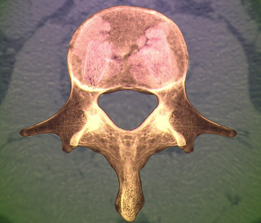
| Coloured composite 2D and 3D computed tomography (CT) axial scan after a vertebral body stenting (VBS) procedure used to treat a horizontal fracture of the L1 vertebra in a 35 year old patient. Both expandable stents (top, pink cages) used for restoring the height of the vertebra and initially stabilising it can be seen. The opaque volume of acrylic cement used to further stabilise and strengthen the vertebra can also be seen. | |
| Lizenzart: | Lizenzpflichtig |
| Credit: | Science Photo Library / Zephyr |
| Bildgröße: | 3969 px × 3402 px |
| Modell-Rechte: | nicht erforderlich |
| Eigentums-Rechte: | nicht erforderlich |
| Restrictions: | - |
Preise für dieses Bild ab 15 €
Universitäten & Organisationen
(Informationsmaterial Digital, Informationsmaterial Print, Lehrmaterial Digital etc.)
ab 15 €
Redaktionell
(Bücher, Bücher: Sach- und Fachliteratur, Digitale Medien (redaktionell) etc.)
ab 30 €
Werbung
(Anzeigen, Aussenwerbung, Digitale Medien, Fernsehwerbung, Karten, Werbemittel, Zeitschriften etc.)
ab 55 €
Handelsprodukte
(bedruckte Textilie, Kalender, Postkarte, Grußkarte, Verpackung etc.)
ab 75 €
Pauschalpreise
Rechtepakete für die unbeschränkte Bildnutzung in Print oder Online
ab 495 €