Oesophageal varices
Bildnummer 13435438
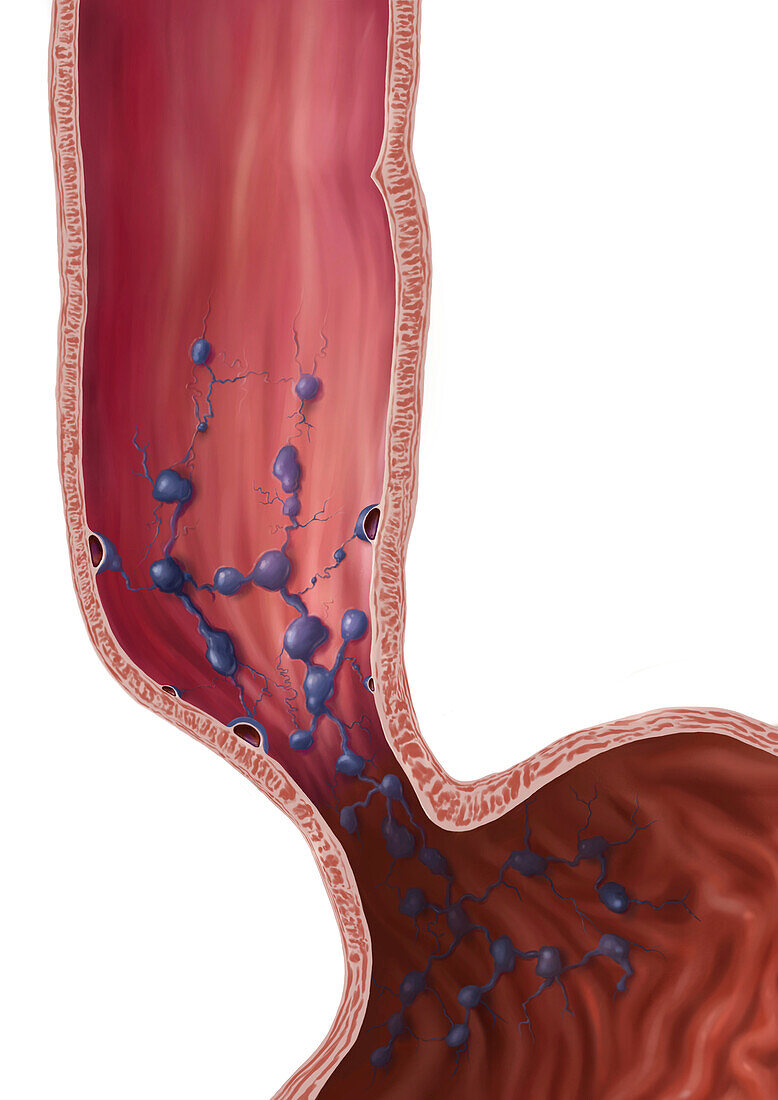
| Illustration of an oesophagus affected by varices (dilated blood vessels, blue) due to liver cirrhosis. Here, the veins in the wall of the oesophagus are dilated. Oesophageal varices are extremely dilated sub-mucosal veins in the lower third of the oesophagus. They are most often a consequence of portal hypertension, high blood pressure in the portal vein and its tributaries. | |
| Lizenzart: | Lizenzpflichtig |
| Credit: | Science Photo Library / MICHAEL HOFFMANN / MEDICAL GRAPHICS |
| Bildgröße: | 2745 px × 3883 px |
| Modell-Rechte: | nicht erforderlich |
| Eigentums-Rechte: | nicht erforderlich |
| Restrictions: | - |
Preise für dieses Bild ab 15 €
Universitäten & Organisationen
(Informationsmaterial Digital, Informationsmaterial Print, Lehrmaterial Digital etc.)
ab 15 €
Redaktionell
(Bücher, Bücher: Sach- und Fachliteratur, Digitale Medien (redaktionell) etc.)
ab 30 €
Werbung
(Anzeigen, Aussenwerbung, Digitale Medien, Fernsehwerbung, Karten, Werbemittel, Zeitschriften etc.)
ab 55 €
Handelsprodukte
(bedruckte Textilie, Kalender, Postkarte, Grußkarte, Verpackung etc.)
ab 75 €
Pauschalpreise
Rechtepakete für die unbeschränkte Bildnutzung in Print oder Online
ab 495 €
Keywords
- abnormal,
- Anatomie,
- anatomisch,
- Biologie,
- biologisch,
- Blutgefäß,
- Bluthochdruck,
- Blutkreislauf,
- Darm,
- Eingeweide,
- Gastroenterologie,
- gastroenterologisch,
- Gefäße,
- Gesundheitswesen,
- GI tract,
- Illustration,
- intern,
- Kondition,
- Krampfadern,
- Kreislauf,
- Kunstwerk,
- Magen-Darm-Trakt,
- Medizin,
- medizinisch,
- menschlicher Körper,
- Niemand,
- Oesophagus,
- Organ,
- Speiseröhre,
- Störung,
- ungesund,
- vaskulär,
- Vene,
- Venen,
- venös,
- Verdauungskanal,
- Verdauungssystem