Heart, illustration
Bildnummer 13386351
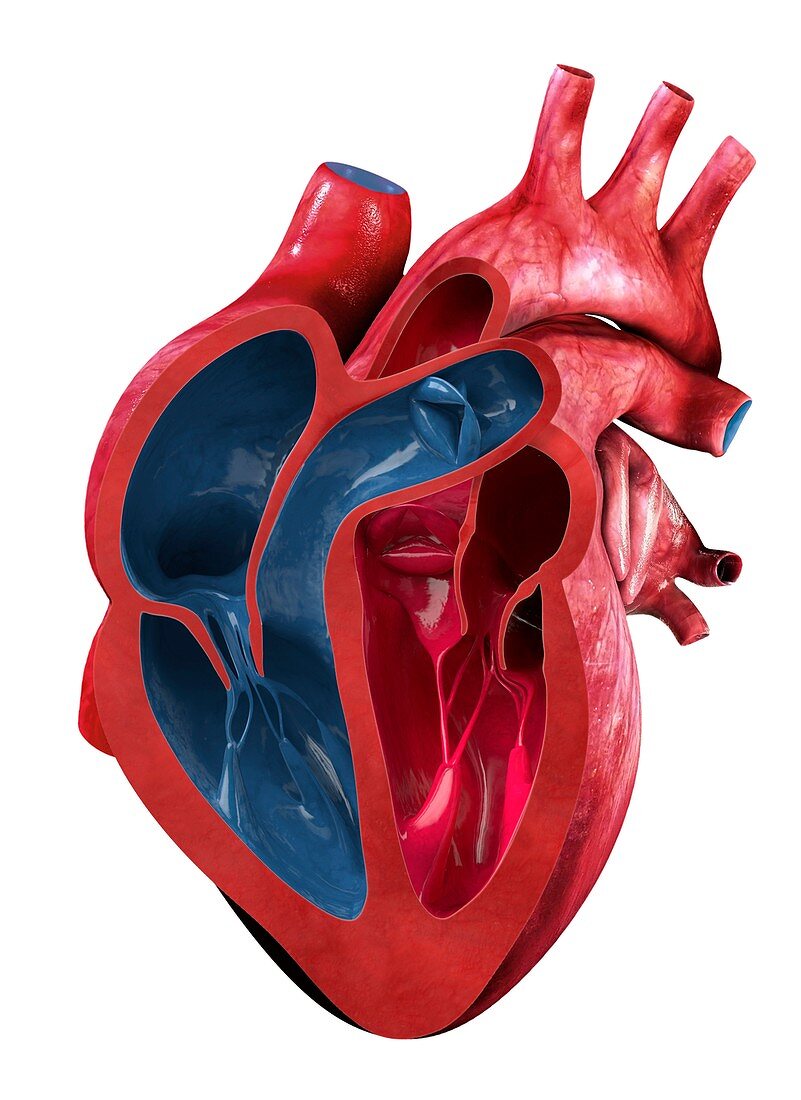
| Illustration of a section through a human heart, showing its internal anatomy. The internal chambers of the heart consist of the left (lower right) and right (bottom left) ventricles, which are separated from the left atrium (upper right) and right atrium (centre-left) by the mitral and tricuspid valves respectively. Deoxygenated blood enters via the vena cava vein (top left) and is pumped into the right atrium (left). Blood leaves the left ventricle (right) for the lungs via the pulmonary artery (top right). Oxygenated blood leaves the heart via the aorta. | |
| Lizenzart: | Lizenzpflichtig |
| Credit: | Science Photo Library / Lunau, Claus |
| Bildgröße: | 3584 px × 4924 px |
| Modell-Rechte: | nicht erforderlich |
| Eigentums-Rechte: | nicht erforderlich |
| Restrictions: | - |
Preise für dieses Bild ab 15 €
Universitäten & Organisationen
(Informationsmaterial Digital, Informationsmaterial Print, Lehrmaterial Digital etc.)
ab 15 €
Redaktionell
(Bücher, Bücher: Sach- und Fachliteratur, Digitale Medien (redaktionell) etc.)
ab 30 €
Werbung
(Anzeigen, Aussenwerbung, Digitale Medien, Fernsehwerbung, Karten, Werbemittel, Zeitschriften etc.)
ab 55 €
Handelsprodukte
(bedruckte Textilie, Kalender, Postkarte, Grußkarte, Verpackung etc.)
ab 75 €
Pauschalpreise
Rechtepakete für die unbeschränkte Bildnutzung in Print oder Online
ab 495 €
Keywords
- Anatomie,
- anatomisch,
- Aorta,
- atrioventrikulär,
- Atrium,
- ausgeschnitten,
- Ausschnitte,
- Biologie,
- biologisch,
- Blutgefäß,
- Gefäße,
- gesund,
- Herz,
- Illustration,
- Kammer,
- kardiovaskular,
- Kreislauf,
- Kunstwerk,
- Lungenarterie,
- Muskel,
- Niemand,
- normal,
- Organ,
- Pulmonalklappe,
- Vena-cava,
- Vene,
- Ventil,
- Ventrikel,
- weißer Hintergrund