Knee anatomy, illustration
Bildnummer 13377701
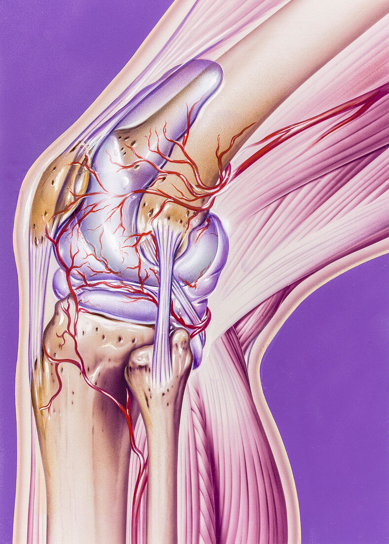
| Knee anatomy, illustration. This is a view of the outside of the knee. The knee joint is formed by the articulation of the femur (thigh bone, top) with the tibia (shin bone, bottom left). The surfaces of the joints are covered by menisci, a form of hyaline cartilage, that act as shock absorbers for the knee protecting the bone surface from wear and tear. The patella (kneecap, brown) is at left. The patella tendon attaches the bottom of the patella to the tibia. The fibular collateral ligament (FCL) connects the femur and fibula (calf bone, bottom centre). | |
| Lizenzart: | Lizenzpflichtig |
| Credit: | Science Photo Library / Bavosi, John |
| Bildgröße: | 4528 px × 6323 px |
| Modell-Rechte: | nicht erforderlich |
| Eigentums-Rechte: | nicht erforderlich |
| Restrictions: | - |
Preise für dieses Bild ab 15 €
Universitäten & Organisationen
(Informationsmaterial Digital, Informationsmaterial Print, Lehrmaterial Digital etc.)
ab 15 €
Redaktionell
(Bücher, Bücher: Sach- und Fachliteratur, Digitale Medien (redaktionell) etc.)
ab 30 €
Werbung
(Anzeigen, Aussenwerbung, Digitale Medien, Fernsehwerbung, Karten, Werbemittel, Zeitschriften etc.)
ab 55 €
Handelsprodukte
(bedruckte Textilie, Kalender, Postkarte, Grußkarte, Verpackung etc.)
ab 75 €
Pauschalpreise
Rechtepakete für die unbeschränkte Bildnutzung in Print oder Online
ab 495 €
Keywords
- Anatomie,
- anatomisch,
- Bein,
- Biologie,
- biologisch,
- Blutgefäße,
- Blutversorgung,
- draußen,
- Femur,
- Gelenk,
- gesund,
- Illustration,
- Joint,
- Knie,
- Kniescheibe,
- Knochen,
- Kunstwerk,
- Lila Hintergrund,
- menschlicher Körper,
- Niemand,
- normal,
- Oberfläche,
- Oberschenkelknochen,
- Profil,
- Schienbein,
- Seitenansicht,
- Wadenbein