Haemangioblastoma, light micrograph
Bildnummer 13296463
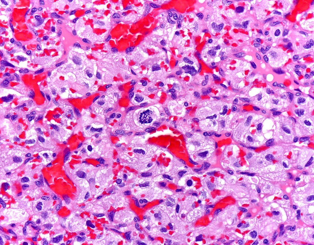
| Haemangioblastoma, light micrograph. Hemangioblastoma is a WHO Grade I tumour that may occur sporadically or in the setting of von Hippel Lindau disease (about 25% of patients). The peak incidence is seen in the 3rd and 4th decades. The most common location is cerebellum. On MRI with contrast, the tumour usually appears a sharply demarcated enhancing mass with cystic areas in the cerebellum. This appearance is seen in about 60% of cases; remaining 40% of cases are entirely solid. About 10-25% of patients with hemangioblastoma carry a germline mutation in the VHL gene. Grossly, hemangioblastomas are generally well circumscribed with solid and cystic components. They are usually dark red-brown given extensive vascularity. This high magnification image of a hemangioblastoma shows stromal cells with abundant vacuolated or lightly eosinophilic cytoplasm. Mitotic activity is generally not increased; but occasional mitotic figures may be present (as shown here). Necrosis is not a prominent feature. | |
| Lizenzart: | Lizenzpflichtig |
| Credit: | Science Photo Library / WEBPATHOLOGY |
| Bildgröße: | 4096 px × 3200 px |
| Modell-Rechte: | nicht erforderlich |
| Eigentums-Rechte: | nicht erforderlich |
| Restrictions: | - |
Preise für dieses Bild ab 15 €
Universitäten & Organisationen
(Informationsmaterial Digital, Informationsmaterial Print, Lehrmaterial Digital etc.)
ab 15 €
Redaktionell
(Bücher, Bücher: Sach- und Fachliteratur, Digitale Medien (redaktionell) etc.)
ab 30 €
Werbung
(Anzeigen, Aussenwerbung, Digitale Medien, Fernsehwerbung, Karten, Werbemittel, Zeitschriften etc.)
ab 55 €
Handelsprodukte
(bedruckte Textilie, Kalender, Postkarte, Grußkarte, Verpackung etc.)
ab 75 €
Pauschalpreise
Rechtepakete für die unbeschränkte Bildnutzung in Print oder Online
ab 495 €
Keywords
- abnormal,
- Epilepsie,
- Gehirn,
- Hämangioblastom,
- Histologie,
- histologisch,
- Histopathologie,
- histopathologisch,
- Karzinom,
- Kondition,
- Krankheit,
- Krebs,
- krebsartig,
- Lichtmikroskop,
- lichtmikroskopische Aufnahme,
- maligne,
- Malignom,
- Neuroglia,
- Neurologie,
- neurologisch,
- Niemand,
- Onkologie,
- Pathologie,
- pathologisch,
- Störung,
- Tumor,
- ungesund,
- zentrales Nervensystem