Central neurocytoma, light micrograph
Bildnummer 13296440
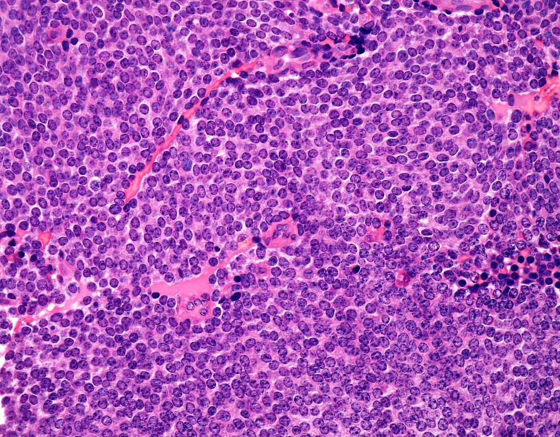
| Central neurocytoma, light micrograph. Central neurocytomas are considered to be WHO Grade II lesions with good prognosis. The peak incidence is between 3rd and 5th decades. They are usually intraventricular and most often arise in the lateral ventricles near the foramen of Monro. Symptoms are related to increased intracranial pressure. Central neurocytomas are composed of a uniform population of round cells with scant cytoplasm in a fibrillary background. The nuclei have finely speckled chromatin and punctate nucleoli. A delicate vascular network of capillaries is usually present. The tumour cells show evidence of neural differentiation. | |
| Lizenzart: | Lizenzpflichtig |
| Credit: | Science Photo Library / WEBPATHOLOGY |
| Bildgröße: | 4096 px × 3200 px |
| Modell-Rechte: | nicht erforderlich |
| Eigentums-Rechte: | nicht erforderlich |
| Restrictions: | - |
Preise für dieses Bild ab 15 €
Universitäten & Organisationen
(Informationsmaterial Digital, Informationsmaterial Print, Lehrmaterial Digital etc.)
ab 15 €
Redaktionell
(Bücher, Bücher: Sach- und Fachliteratur, Digitale Medien (redaktionell) etc.)
ab 30 €
Werbung
(Anzeigen, Aussenwerbung, Digitale Medien, Fernsehwerbung, Karten, Werbemittel, Zeitschriften etc.)
ab 55 €
Handelsprodukte
(bedruckte Textilie, Kalender, Postkarte, Grußkarte, Verpackung etc.)
ab 75 €
Pauschalpreise
Rechtepakete für die unbeschränkte Bildnutzung in Print oder Online
ab 495 €
Keywords
- abnormal,
- Epilepsie,
- Gehirn,
- Histologie,
- histologisch,
- Histopathologie,
- histopathologisch,
- Karzinom,
- Kondition,
- Krankheit,
- Krebs,
- krebsartig,
- Lichtmikroskop,
- lichtmikroskopische Aufnahme,
- maligne,
- Malignom,
- neural,
- Neurologie,
- neurologisch,
- Niemand,
- Onkologie,
- Pathologie,
- pathologisch,
- Störung,
- Tumor,
- ungesund,
- zentrales Nervensystem