Diffuse astrocytoma, light micrograph
Bildnummer 13296339
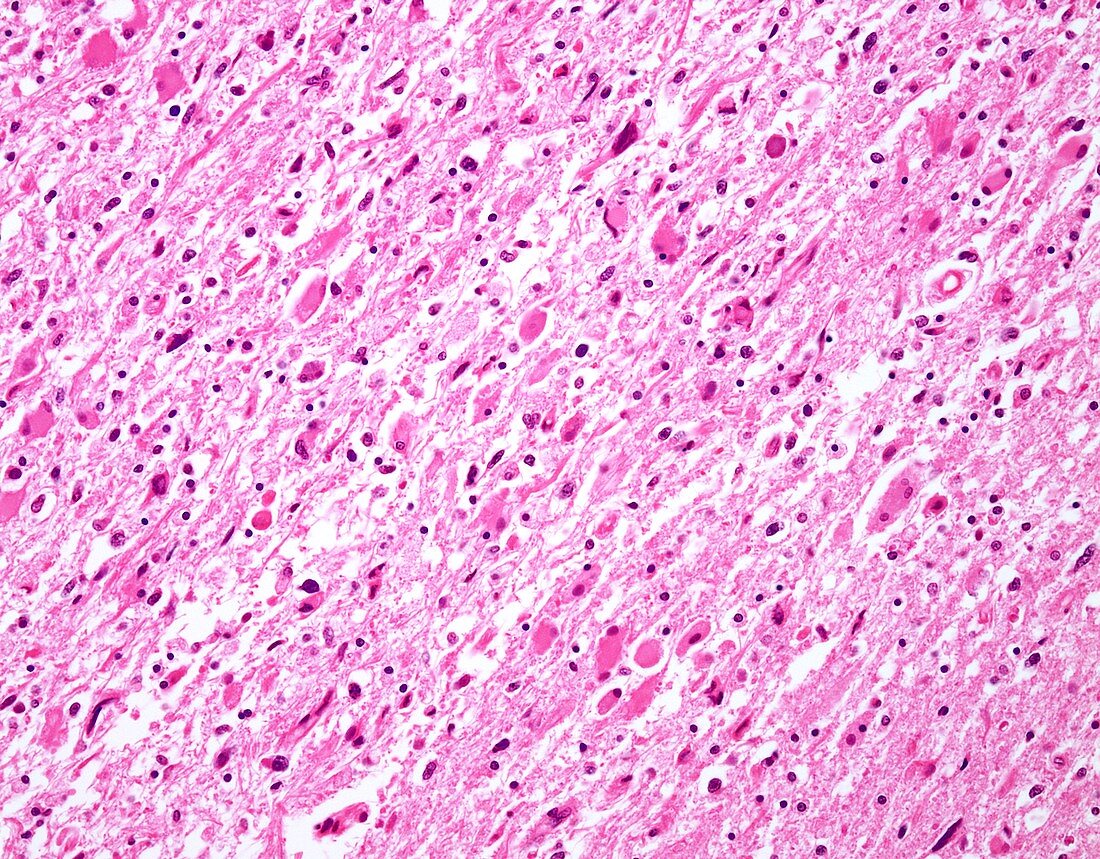
| Diffuse astrocytoma, light micrograph. Infiltrating astrocytomas account for almost 80% of adult primary brain tumours. The usual location is cerebral hemispheres and the most commonly affected age group is 4th to 6th decades. The histologic spectrum ranges from diffuse fibrillary astrocytomas (WHO Grade II; shown in this image), through anaplastic astrocytomas (WHO Grade III) to Glioblastoma (WHO Grade IV). WHO Grade I is reserved for Pilocytic astrocytomas. This image of diffuse astrocytoma (WHO Grade II) shows increased number of glial cells (cells with copious eosinophilic cytoplasm), mild nuclear pleomorphism, and a network of delicate astrocytic processes giving a fibrillary appearance to the background. | |
| Lizenzart: | Lizenzpflichtig |
| Credit: | Science Photo Library / WEBPATHOLOGY |
| Bildgröße: | 4096 px × 3200 px |
| Modell-Rechte: | nicht erforderlich |
| Eigentums-Rechte: | nicht erforderlich |
| Restrictions: | - |
Preise für dieses Bild ab 15 €
Universitäten & Organisationen
(Informationsmaterial Digital, Informationsmaterial Print, Lehrmaterial Digital etc.)
ab 15 €
Redaktionell
(Bücher, Bücher: Sach- und Fachliteratur, Digitale Medien (redaktionell) etc.)
ab 30 €
Werbung
(Anzeigen, Aussenwerbung, Digitale Medien, Fernsehwerbung, Karten, Werbemittel, Zeitschriften etc.)
ab 55 €
Handelsprodukte
(bedruckte Textilie, Kalender, Postkarte, Grußkarte, Verpackung etc.)
ab 75 €
Pauschalpreise
Rechtepakete für die unbeschränkte Bildnutzung in Print oder Online
ab 495 €
Keywords
- abnormal,
- Astrozyten,
- Epilepsie,
- Gehirn,
- Gliom,
- Histologie,
- histologisch,
- Histopathologie,
- histopathologisch,
- Idh,
- Idh-Mutant,
- IDH-Mutation,
- IDH-Wildtyp,
- Karzinom,
- Kondition,
- Krankheit,
- Krebs,
- krebsartig,
- Lichtmikroskop,
- lichtmikroskopische Aufnahme,
- maligne,
- Malignom,
- Neuroglia,
- Neurologie,
- neurologisch,
- Niemand,
- Onkologie,
- Pathologie,
- pathologisch,
- Störung,
- Tumor,
- ungesund,
- zentrales Nervensystem