Brain activity during a visual language task, fMRI scan
Bildnummer 13258388
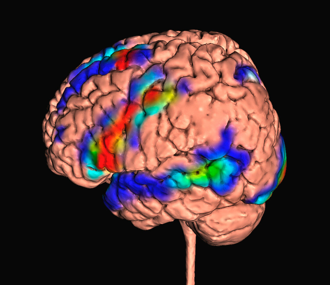
| Composite image of the results of a functional magnetic resonance imaging (fMRI) scan overlaid on a model of the brain. Functional magnetic resonance imaging (fMRI) studies show areas of the brain with increased activity during particular tasks. This scan shows the results of a visual sentence completion task, which activated Broca's area in the left frontal (left) lobe and Wernicke's area in the left temporal lobe. Broca's area is linked to speech production, while Wernicke's area is involved in the comprehension of written and spoken language. Areas of the visual cortex at the rear (right) of the brain are also activated. | |
| Lizenzart: | Lizenzpflichtig |
| Credit: | Science Photo Library / Fung, K.H. |
| Bildgröße: | 9000 px × 7788 px |
| Modell-Rechte: | nicht erforderlich |
| Eigentums-Rechte: | nicht erforderlich |
| Restrictions: | - |
Preise für dieses Bild ab 15 €
Universitäten & Organisationen
(Informationsmaterial Digital, Informationsmaterial Print, Lehrmaterial Digital etc.)
ab 15 €
Redaktionell
(Bücher, Bücher: Sach- und Fachliteratur, Digitale Medien (redaktionell) etc.)
ab 30 €
Werbung
(Anzeigen, Aussenwerbung, Digitale Medien, Fernsehwerbung, Karten, Werbemittel, Zeitschriften etc.)
ab 55 €
Handelsprodukte
(bedruckte Textilie, Kalender, Postkarte, Grußkarte, Verpackung etc.)
ab 75 €
Pauschalpreise
Rechtepakete für die unbeschränkte Bildnutzung in Print oder Online
ab 495 €
Keywords
- Anatomie,
- anatomisch,
- ausgeschnitten,
- Ausschnitte,
- Bereiche,
- Biologie,
- biologisch,
- cerebral,
- cgi,
- Cortex,
- digital generiert,
- farbig,
- FMRI,
- Frontal,
- Funktion,
- Funktional,
- gefärbt,
- Gehirn,
- Hinter-,
- Hirnscan,
- Illustration,
- Karte,
- Kunstwerk,
- Lappen,
- Magnetresonanztomografie,
- menschlicher Körper,
- MRT-Untersuchung,
- Neuroimaging,
- Neurologie,
- neurologisch,
- Neurowissenschaften,
- Niemand,
- Organ,
- Rede,
- schwarzer Hintergrund,
- Sprache,
- Zusammengesetzt