Epiphyseal growth plate, light micrograph
Bildnummer 13243165
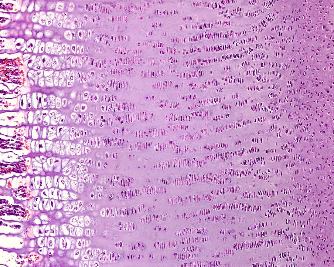
| Light micrograph of the epiphyseal growth plate of a developing long bone. The epiphyseal cartilage shows the following layers, from right to left: resting hyaline cartilage, zone of proliferation, zone of hypertrophy with large lacunae, zone of calcification (not visible as such, but can be identified by the degeneration of the chondrocytes leaving empty cartilaginous lacunae), and an ossification zone of compact appearance, where there is an invasion of blood vessels and osteogenic cells. | |
| Lizenzart: | Lizenzpflichtig |
| Credit: | Science Photo Library / JOSE CALVO |
| Bildgröße: | 3840 px × 3072 px |
| Modell-Rechte: | nicht erforderlich |
| Eigentums-Rechte: | nicht erforderlich |
| Restrictions: | - |
Preise für dieses Bild ab 15 €
Universitäten & Organisationen
(Informationsmaterial Digital, Informationsmaterial Print, Lehrmaterial Digital etc.)
ab 15 €
Redaktionell
(Bücher, Bücher: Sach- und Fachliteratur, Digitale Medien (redaktionell) etc.)
ab 30 €
Werbung
(Anzeigen, Aussenwerbung, Digitale Medien, Fernsehwerbung, Karten, Werbemittel, Zeitschriften etc.)
ab 55 €
Handelsprodukte
(bedruckte Textilie, Kalender, Postkarte, Grußkarte, Verpackung etc.)
ab 75 €
Pauschalpreise
Rechtepakete für die unbeschränkte Bildnutzung in Print oder Online
ab 495 €