Herniated disc, CT scan
Bildnummer 13218972
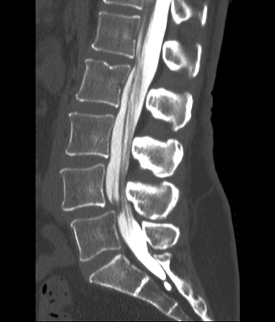
| Computed tomography (CT) scan of the lower back of a 38 year old female patient with sciatica. The spinal cord (white) appears to be compressed (dark area) by a herniated (slipped) intervertebral disc between the fourth and fifth lumbar (lower) vertebra (backbones, blocks down left). This is most likely the cause of the pain. | |
| Lizenzart: | Lizenzpflichtig |
| Credit: | Science Photo Library / Zephyr |
| Bildgröße: | 2442 px × 2862 px |
| Modell-Rechte: | nicht erforderlich |
| Eigentums-Rechte: | nicht erforderlich |
| Restrictions: | - |
Preise für dieses Bild ab 15 €
Universitäten & Organisationen
(Informationsmaterial Digital, Informationsmaterial Print, Lehrmaterial Digital etc.)
ab 15 €
Redaktionell
(Bücher, Bücher: Sach- und Fachliteratur, Digitale Medien (redaktionell) etc.)
ab 30 €
Werbung
(Anzeigen, Aussenwerbung, Digitale Medien, Fernsehwerbung, Karten, Werbemittel, Zeitschriften etc.)
ab 55 €
Handelsprodukte
(bedruckte Textilie, Kalender, Postkarte, Grußkarte, Verpackung etc.)
ab 75 €
Pauschalpreise
Rechtepakete für die unbeschränkte Bildnutzung in Print oder Online
ab 495 €
Keywords
- 30er Jahre,
- abnormal,
- Bandscheiben,
- Computertomographie,
- CT-Scan,
- Diagnose,
- dreißiger Jahre,
- Einfarbig,
- Frau,
- Ischias,
- Kondition,
- Krankheit,
- L4,
- L5,
- Lendenwirbelsäule,
- Medizin,
- medizinisch,
- menschlicher Körper,
- niedriger,
- Niemand,
- Profil,
- Rückenmark,
- Rückgrat,
- Scheibe,
- schwarz und weiß,
- schwarzer Hintergrund,
- Seitenansicht,
- Störung,
- ungesund,
- Weiblich,
- Wirbel,
- Wirbelsäule,
- Wirbelsäulen-,
- zentrales Nervensystem