Skeletal muscle, SEM-TEM comparison
Bildnummer 13218073
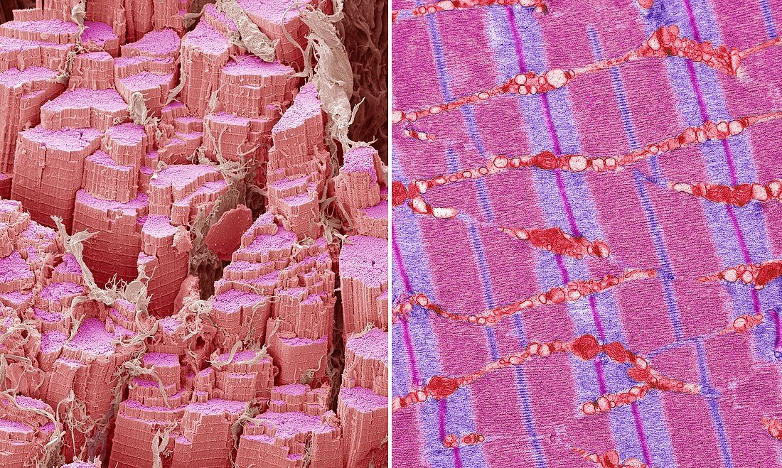
| Skeletal muscle. Comparison between a scanning election micrograph (SEM left) and transmission electron micrograph (TEM right) of skeletal, or striated, muscle. The striations are due to the muscle fibrils, which run left to right. These are made up of the proteins actin and myosin. In the TEM image the lighter regions are stretches of actin, the darker regions are actin and myosin. Muscles contract when actin slides over the myosin. The energy for the contractions comes from mitochondria (orange TEM). Skeletal muscle is under conscious control. Magnification: x SEM 2500, TEM x10000 when printed at 10 centimetres high. | |
| Lizenzart: | Lizenzpflichtig |
| Credit: | Science Photo Library / Gschmeissner, Steve |
| Bildgröße: | 7589 px × 4548 px |
| Modell-Rechte: | nicht erforderlich |
| Restrictions: | - |
Preise für dieses Bild ab 15 €
Universitäten & Organisationen
(Informationsmaterial Digital, Informationsmaterial Print, Lehrmaterial Digital etc.)
ab 15 €
Redaktionell
(Bücher, Bücher: Sach- und Fachliteratur, Digitale Medien (redaktionell) etc.)
ab 30 €
Werbung
(Anzeigen, Aussenwerbung, Digitale Medien, Fernsehwerbung, Karten, Werbemittel, Zeitschriften etc.)
ab 55 €
Handelsprodukte
(bedruckte Textilie, Kalender, Postkarte, Grußkarte, Verpackung etc.)
ab 75 €
Pauschalpreise
Rechtepakete für die unbeschränkte Bildnutzung in Print oder Online
ab 495 €