Lung airways in bronchitis, comparative illustrations
Bildnummer 12971388
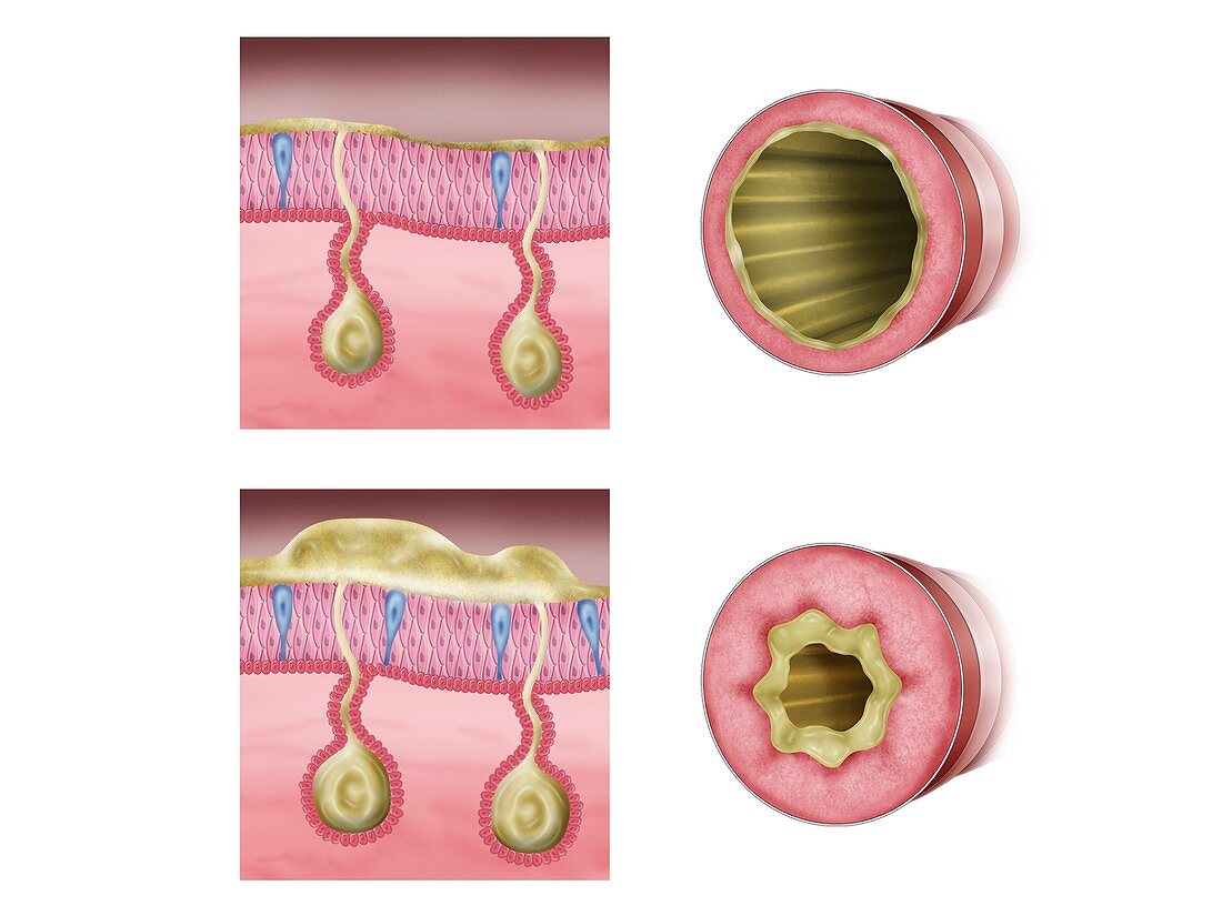
| Lung airways in bronchitis, comparative illustrations. The artworks show cross-sections through a normal airway (top) and an inflamed, constricted airway (bottom). At left, the process of mucus production is shown in vertical section through the airway walls. The airways have a ring of smooth muscle and a thin layer of endothelial tissue. In the inflamed airway, the smooth muscle has contracted, and endothelial cell debris and mucus are partially blocking the airway. This is seen in conditions such as chronic bronchitis. Drugs to treat the condition reduce the inflammation and widen the airways, making it easier to breathe. | |
| Lizenzart: | Lizenzpflichtig |
| Credit: | Science Photo Library / De Angelis, Maurizio |
| Bildgröße: | 4827 px × 3620 px |
| Modell-Rechte: | nicht erforderlich |
| Eigentums-Rechte: | nicht erforderlich |
| Restrictions: | - |
Preise für dieses Bild ab 15 €
Universitäten & Organisationen
(Informationsmaterial Digital, Informationsmaterial Print, Lehrmaterial Digital etc.)
ab 15 €
Redaktionell
(Bücher, Bücher: Sach- und Fachliteratur, Digitale Medien (redaktionell) etc.)
ab 30 €
Werbung
(Anzeigen, Aussenwerbung, Digitale Medien, Fernsehwerbung, Karten, Werbemittel, Zeitschriften etc.)
ab 55 €
Handelsprodukte
(bedruckte Textilie, Kalender, Postkarte, Grußkarte, Verpackung etc.)
ab 75 €
Pauschalpreise
Rechtepakete für die unbeschränkte Bildnutzung in Print oder Online
ab 495 €
Keywords
- Abschnitte,
- Alveolen,
- Anatomie,
- anatomisch,
- Atemweg,
- Atemwege,
- Atmungssystem,
- ausgeschnitten,
- Ausschnitte,
- Bronchiole,
- Bronchiolen,
- Bronchitis,
- Bronchus,
- chronische Bronchitis,
- Cutaway,
- diagonal,
- endothelial,
- Entzündung,
- Illustration,
- Kondition,
- Kontraktion,
- Kunstwerk,
- Lumen,
- Lunge,
- Lungen,
- Medizin,
- medizinisch,
- menschlicher Körper,
- Niemand,
- Pathologie,
- pathologisch,
- Querschnitt,
- Querschnitte,
- Schicht,
- Schichten,
- Schleim,
- Sektion,
- sektioniert,
- Störung,
- Trümmer,
- Vergleich,
- vergleichen,
- verstopft,
- weißer Hintergrund