Inner ear anatomy, illustration
Bildnummer 12971323
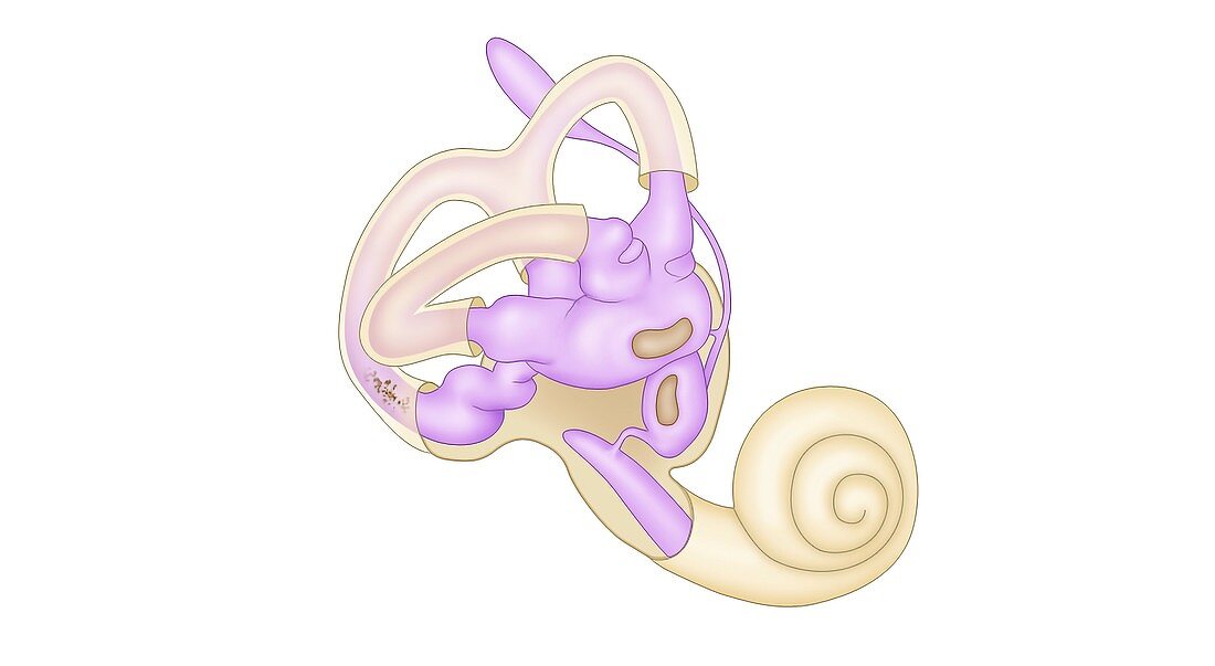
| Inner ear anatomy. Illustration of the anatomy of the sensory organs of the human inner ear. The inner ear contains a maze of fluid-filled passages called the labyrinth (pink). The cochlea (lower right) is a hollow spiral that contains microscopic hairs that respond to sound vibrations from the middle ear (not seen). Nerves connected to the cochlea carry the sound information as electric impulses to the brain. The three loops at upper left are the semi-circular canals. These provide the sense of balance by detecting movement of fluid within them. At centre is the vestibule of the ear, which includes structures called the utricle and saccule. These are the otolith organs, and also detect motion. | |
| Lizenzart: | Lizenzpflichtig |
| Credit: | Science Photo Library / De Angelis, Maurizio |
| Bildgröße: | 4000 px × 2194 px |
| Modell-Rechte: | nicht erforderlich |
| Eigentums-Rechte: | nicht erforderlich |
| Restrictions: | - |
Preise für dieses Bild ab 15 €
Universitäten & Organisationen
(Informationsmaterial Digital, Informationsmaterial Print, Lehrmaterial Digital etc.)
ab 15 €
Redaktionell
(Bücher, Bücher: Sach- und Fachliteratur, Digitale Medien (redaktionell) etc.)
ab 30 €
Werbung
(Anzeigen, Aussenwerbung, Digitale Medien, Fernsehwerbung, Karten, Werbemittel, Zeitschriften etc.)
ab 55 €
Handelsprodukte
(bedruckte Textilie, Kalender, Postkarte, Grußkarte, Verpackung etc.)
ab 75 €
Pauschalpreise
Rechtepakete für die unbeschränkte Bildnutzung in Print oder Online
ab 495 €