Distal finger joint anatomy, illustration
Bildnummer 12971283
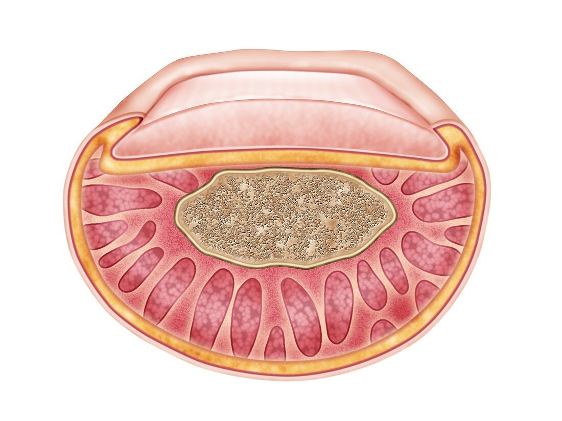
| Distal finger joint anatomy, illustration. Cross-section through the distal portion of a finger. The distal part of a fnger is the final portion, the end that is distant from the point of attachment. The section has passed through the fingernail, and tissue layers including skin, fat, connective tissue, muscle and bone. The bone here is the distal phalanx. | |
| Lizenzart: | Lizenzpflichtig |
| Credit: | Science Photo Library / De Angelis, Maurizio |
| Bildgröße: | 4831 px × 3617 px |
| Modell-Rechte: | nicht erforderlich |
| Eigentums-Rechte: | nicht erforderlich |
| Restrictions: | - |
Preise für dieses Bild ab 15 €
Universitäten & Organisationen
(Informationsmaterial Digital, Informationsmaterial Print, Lehrmaterial Digital etc.)
ab 15 €
Redaktionell
(Bücher, Bücher: Sach- und Fachliteratur, Digitale Medien (redaktionell) etc.)
ab 30 €
Werbung
(Anzeigen, Aussenwerbung, Digitale Medien, Fernsehwerbung, Karten, Werbemittel, Zeitschriften etc.)
ab 55 €
Handelsprodukte
(bedruckte Textilie, Kalender, Postkarte, Grußkarte, Verpackung etc.)
ab 75 €
Pauschalpreise
Rechtepakete für die unbeschränkte Bildnutzung in Print oder Online
ab 495 €