Lung alveoli anatomy, 19th century
Bildnummer 12970454
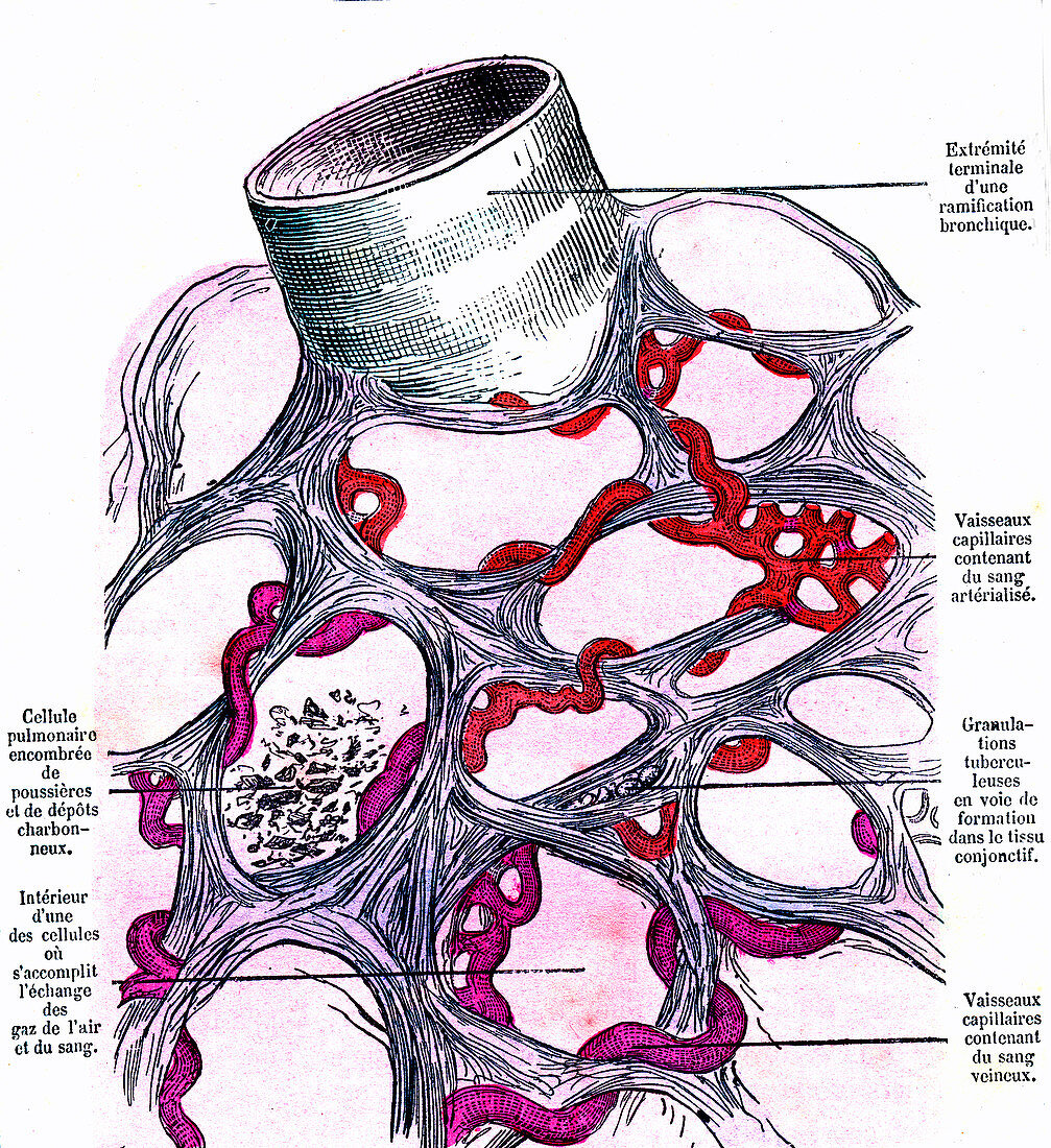
| Lung alveoli anatomy, 19th-century illustration. At upper left is part of a terminal bronchiole, which ends in the alveolar sacs shown below in cross-section. Venous (purple) and arterial (red) capillaries are shown meshed in the alveoli, which are the site of gas exchange in the lungs. Carbon dioxide and oxygen are the gases exchanged here. Also shown are deposits of carbon (lower left), and tuberculosis deposits (tubercules, centre). The labels are in French. Published in 'La Vie Normale et la Sante' (Normal Life and Health, Paris, 1881) by Dr Jules Rengade. | |
| Lizenzart: | Lizenzpflichtig |
| Credit: | Science Photo Library / Collection Abecasis |
| Bildgröße: | 2963 px × 3236 px |
| Modell-Rechte: | nicht erforderlich |
| Eigentums-Rechte: | nicht erforderlich |
| Restrictions: | - |
Preise für dieses Bild ab 15 €
Universitäten & Organisationen
(Informationsmaterial Digital, Informationsmaterial Print, Lehrmaterial Digital etc.)
ab 15 €
Redaktionell
(Bücher, Bücher: Sach- und Fachliteratur, Digitale Medien (redaktionell) etc.)
ab 30 €
Werbung
(Anzeigen, Aussenwerbung, Digitale Medien, Fernsehwerbung, Karten, Werbemittel, Zeitschriften etc.)
ab 55 €
Handelsprodukte
(bedruckte Textilie, Kalender, Postkarte, Grußkarte, Verpackung etc.)
ab 75 €
Pauschalpreise
Rechtepakete für die unbeschränkte Bildnutzung in Print oder Online
ab 495 €
Keywords
- 1800er Jahre,
- 19. Jahrhundert,
- abnormal,
- Alveole,
- Alveolen,
- Anatomie,
- anatomisch,
- Atemweg,
- Atemwege,
- beschriftet,
- Biologie,
- biologisch,
- Blutgefäße,
- Etikette,
- Etiketten,
- europäisch,
- Französisch,
- Französisch Sprache,
- Geschichte,
- gesund,
- historisch,
- Illustration,
- Kapillarbett,
- Kohlenstoff,
- Kondition,
- Krankheit,
- Kunstwerk,
- Lunge,
- Lungen,
- Medizin,
- medizinisch,
- menschlicher Körper,
- Niemand,
- normal,
- Organ,
- Partikel,
- pulmonal,
- Störung,
- Text,
- Truhe,
- Tuberkulose,
- ungesund,
- vaskulär,
- Verschmutzung