Fruit fly leg, SEM
Bildnummer 12968945
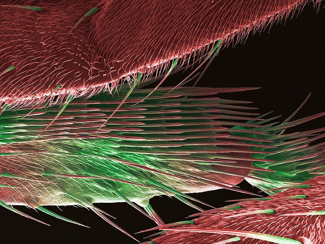
| Close up of the leg of a fruit fly (Drosophila melanogaster), coloured scanning electron micrograph (SEM). Limb or appendage development in vertebrates and invertebrates has been of interest to researchers for many years with Drosophila leg development proving to be a good model system. It has helped researchers gain an insight into developmental processes required for tissue patterning such as movement and shape changes of cells, localized cell growth and proliferation and sub division of the tissue into distinct parts according to positional information. Magnification: x300 when printed at 10cm wide | |
| Lizenzart: | Lizenzpflichtig |
| Credit: | Science Photo Library / Anne Weston, EM STP, the Francis Crick Institute |
| Bildgröße: | 4840 px × 3643 px |
| Modell-Rechte: | nicht erforderlich |
| Eigentums-Rechte: | nicht erforderlich |
| Restrictions: |
|
Preise für dieses Bild ab 15 €
Universitäten & Organisationen
(Informationsmaterial Digital, Informationsmaterial Print, Lehrmaterial Digital etc.)
ab 15 €
Redaktionell
(Bücher, Bücher: Sach- und Fachliteratur, Digitale Medien (redaktionell) etc.)
ab 30 €
Werbung
(Anzeigen, Aussenwerbung, Digitale Medien, Fernsehwerbung, Karten, Werbemittel, Zeitschriften etc.)
ab 55 €
Handelsprodukte
(bedruckte Textilie, Kalender, Postkarte, Grußkarte, Verpackung etc.)
ab 75 €
Pauschalpreise
Rechtepakete für die unbeschränkte Bildnutzung in Print oder Online
ab 495 €
Keywords
- Bein,
- Biologie,
- biologisch,
- Drosophila melanogaster,
- Entomologie,
- entomologisch,
- Entwicklungsbiologie,
- entwicklungsgemäß,
- experimentell,
- farbig,
- Fauna,
- Fliege,
- Fruchtfliege,
- gefärbt,
- Genetik,
- Genom,
- Glied,
- Insekt,
- Kondition,
- Krankheit,
- Natur,
- Rasterelektronenmikroskopie,
- rasterelektronenmikroskopische Aufnahme,
- REM,
- Störung,
- Tier,
- Tierwelt,
- Zoologie,
- zoologisch