Trachea, SEM-TEM comparison
Bildnummer 12948543
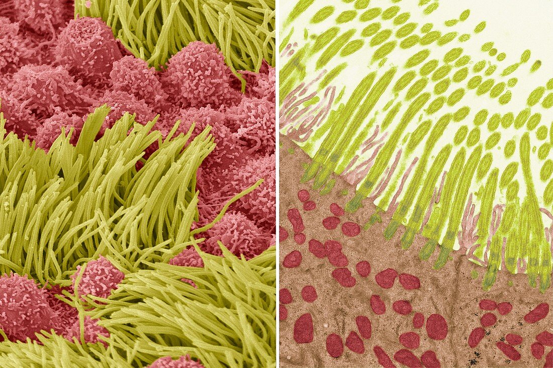
| Comparison between a transmission electron micrograph (TEM, right) and scanning electron micrograph (SEM, left) of tracheal epithelium. In both images cilia are coloured green while the microvilli of the mucous secreting goblet cells are red. In the TEM the internal structure of the cilia is visible; the mitochondria are seen in red within the cell. Mucous serves to trap tiny foreign particles in inhaled air which are transported by the movement of the cilia upwards and out of the respiratory tract, keeping the lungs and airways clear. In the SEM the surface structure of the specimen is visible. Magnification SEM X5000, TEM X9000 when printed at 10cm high. For a series of comparisons between SEMs and TEMs see images C047/7006 to C047/7034. | |
| Lizenzart: | Lizenzpflichtig |
| Credit: | Science Photo Library / Gschmeissner, Steve |
| Bildgröße: | 6828 px × 4542 px |
| Modell-Rechte: | nicht erforderlich |
| Eigentums-Rechte: | nicht erforderlich |
| Restrictions: | - |
Preise für dieses Bild ab 15 €
Universitäten & Organisationen
(Informationsmaterial Digital, Informationsmaterial Print, Lehrmaterial Digital etc.)
ab 15 €
Redaktionell
(Bücher, Bücher: Sach- und Fachliteratur, Digitale Medien (redaktionell) etc.)
ab 30 €
Werbung
(Anzeigen, Aussenwerbung, Digitale Medien, Fernsehwerbung, Karten, Werbemittel, Zeitschriften etc.)
ab 55 €
Handelsprodukte
(bedruckte Textilie, Kalender, Postkarte, Grußkarte, Verpackung etc.)
ab 75 €
Pauschalpreise
Rechtepakete für die unbeschränkte Bildnutzung in Print oder Online
ab 495 €
Keywords
- Biologie,
- biologisch,
- Farbig,
- gefärbt,
- gesund,
- Kultur,
- Luftröhre,
- menschlicher Körper,
- Mikroskop,
- Mikrovilli,
- Mitochondrien,
- normal,
- rasterelektronenmikroskopische Aufnahme,
- Reihenfolge,
- REM,
- Schleim,
- Serie,
- Struktur,
- tem,
- Transmissionselektronenmikroskopie,
- transmissionselektronenmikroskopische Aufnahme,
- Vergleich,
- vergleichen,
- verglichen,
- Wimpern,
- Zelle